



Cardiac Amyloidosis: Symptoms, Red Flags & Diagnosis
Cardiac amyloidosis is an infiltrative cardiomyopathy caused by the deposition of amyloid fibrils within the heart. Progressive amyloid deposition alters myocardial structure and function and can lead to heart failure, arrhythmias, conduction disease, and other cardiovascular complications. The two major forms encountered in clinical practice are transthyretin cardiac amyloidosis (ATTR-CM) and immunoglobulin light-chain cardiac amyloidosis (AL-CM) (1, 2).
Cardiac amyloidosis has historically been under-recognized because its manifestations overlap with more common cardiovascular conditions. Greater awareness of characteristic cardiac and extracardiac red flags, together with modern imaging and laboratory testing, now allows many patients to be identified without relying solely on endomyocardial biopsy (1, 2).
What Is Cardiac Amyloidosis?
Amyloidosis occurs when normally soluble precursor proteins misfold and aggregate into insoluble amyloid fibrils. When these fibrils accumulate within the myocardium and other cardiac structures, they can increase ventricular wall thickness, impair relaxation, alter myocardial deformation and eventually produce progressive cardiac dysfunction (1, 2).
Although several amyloid proteins can involve the heart, the overwhelming majority of clinically important cardiac amyloidosis is caused by either transthyretin or immunoglobulin light chains.
The two major types of cardiac amyloidosis
- ATTR-CM: caused by misfolded transthyretin and occurring as wild-type ATTR (ATTRwt) or variant/hereditary ATTR (ATTRv).
- AL-CM: caused by amyloid-forming immunoglobulin light chains produced by an abnormal plasma-cell or other B-cell clone.
ATTR-CM and AL-CM: Why the Distinction Matters
ATTR-CM and AL-CM can produce similar cardiac phenotypes, but they arise from fundamentally different diseases and require very different treatment strategies (1, 2).
Transthyretin Cardiac Amyloidosis (ATTR-CM)
Transthyretin is produced predominantly by the liver. In ATTR amyloidosis, transthyretin becomes unstable, misfolds and forms amyloid fibrils that can accumulate in the heart and other tissues.
ATTR-CM occurs in two principal forms (1):
- Wild-type ATTR (ATTRwt): occurs without a pathogenic TTR variant and predominantly affects older adults.
- Variant ATTR (ATTRv): results from a pathogenic or likely pathogenic variant in the TTR gene and can produce cardiac, neurological or mixed phenotypes.
Immunoglobulin Light-Chain Cardiac Amyloidosis (AL-CM)
AL amyloidosis results from an abnormal clonal population of plasma cells or, less commonly, another B-cell disorder producing amyloidogenic immunoglobulin light chains. The heart is frequently involved, and cardiac involvement is a major determinant of prognosis (3).
Clinical priority: AL amyloidosis is a hematologic disease that can progress rapidly. When cardiac amyloidosis is suspected, evaluation for a monoclonal protein should not be delayed while waiting for ATTR-specific investigations.
Why Is Cardiac Amyloidosis Often Missed?
The clinical presentation of cardiac amyloidosis is heterogeneous and frequently resembles more common cardiovascular disorders. Patients may initially be labelled as having hypertensive heart disease, heart failure with preserved ejection fraction (HFpEF), hypertrophic cardiomyopathy, valvular heart disease or nonspecific age-related cardiac dysfunction (1, 2).
No single symptom, ECG finding or echocardiographic feature is sufficiently sensitive and specific to identify every patient. Diagnosis therefore depends on recognizing patterns of findings across the clinical history, ECG, imaging, laboratory tests and extracardiac manifestations.
What Are the Symptoms of Cardiac Amyloidosis?
Symptoms usually reflect heart failure, arrhythmias, conduction disease or systemic amyloid involvement. Common cardiac manifestations include (1, 2):
- Progressive shortness of breath
- Reduced exercise tolerance
- Fatigue
- Peripheral oedema
- Abdominal congestion or early satiety
- Palpitations
- Dizziness or light-headedness
- Syncope or presyncope
These symptoms are nonspecific. Suspicion increases when they occur together with characteristic imaging abnormalities, conduction disease or extracardiac manifestations of systemic amyloidosis.
Cardiac Red Flags
A red flag is a finding that should increase clinical suspicion but does not establish the diagnosis by itself. Current expert guidance emphasizes combining multiple cardiac and extracardiac clues (1, 2).
Important cardiac red flags
- Unexplained increased left ventricular wall thickness
- Heart failure, particularly HFpEF, with otherwise unexplained ventricular thickening
- Relative apical sparing on longitudinal strain imaging
- Disproportionately low ECG voltage relative to ventricular wall thickness
- Atrial fibrillation or other atrial arrhythmias
- Conduction disease or requirement for a pacemaker
- Characteristic cardiac magnetic resonance abnormalities
- Persistent elevation of cardiac biomarkers in an appropriate clinical context
Increased Ventricular Wall Thickness
Increased left ventricular wall thickness on echocardiography is one of the most important clues to cardiac amyloidosis. Unlike true myocardial hypertrophy, the apparent thickening in amyloidosis reflects myocardial infiltration by amyloid and associated tissue expansion (2, 4).
Cardiac amyloidosis should therefore enter the differential diagnosis when increased ventricular wall thickness cannot be adequately explained by hypertension, valvular disease or another established cause.
Low ECG Voltage: Useful but Not Required
A classic teaching point is the apparent mismatch between increased ventricular wall thickness on echocardiography and relatively low QRS voltage on ECG. This can be a useful clue, but low voltage is not present in all patients with cardiac amyloidosis (1, 2).
Do not exclude cardiac amyloidosis simply because the ECG does not show low voltage.
Extracardiac Red Flags
Cardiac amyloidosis is often part of a multisystem disease. Findings outside the heart may precede cardiac symptoms by years and can provide important diagnostic clues (1, 2).
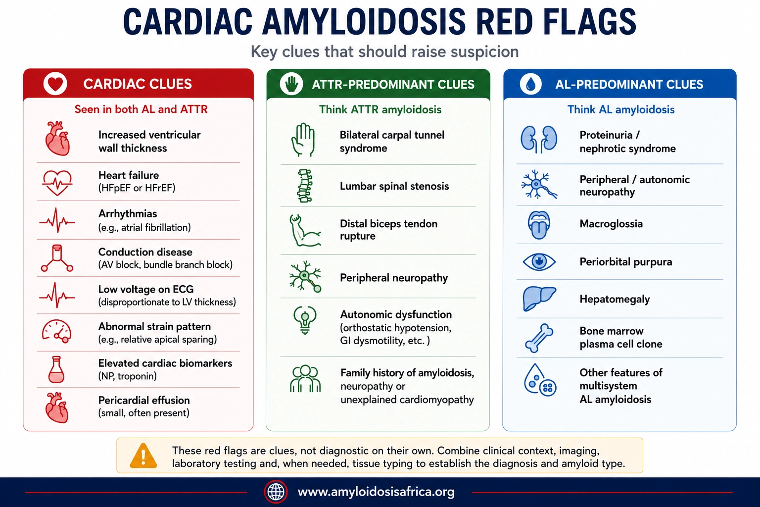
Red Flags That May Suggest ATTR Amyloidosis
- Bilateral carpal tunnel syndrome
- Lumbar spinal stenosis
- Spontaneous distal biceps tendon rupture
- Peripheral neuropathy
- Autonomic dysfunction
- Family history of amyloidosis, neuropathy or unexplained cardiomyopathy
These conditions are common in the general population and are not diagnostic of ATTR amyloidosis by themselves. Their diagnostic value is greatest when they occur in combination with a compatible cardiac phenotype (1).
Red Flags That May Suggest AL Amyloidosis
- Proteinuria or nephrotic syndrome
- Peripheral or autonomic neuropathy
- Macroglossia
- Periorbital purpura
- Unexplained hepatomegaly
- Other manifestations of multisystem light-chain amyloidosis
Macroglossia and periorbital purpura are particularly suggestive when present, although they occur in only a minority of patients (3).
What Can the ECG Show?
The ECG in cardiac amyloidosis may show a range of abnormalities. No individual ECG feature is sufficiently sensitive to rule the disease in or out (1, 4).
Potential findings include:
- Low QRS voltage
- Pseudoinfarction patterns
- Atrial fibrillation
- Atrioventricular conduction abnormalities
- Bundle branch block
- Other conduction disturbances
The ECG is therefore best interpreted as one component of a multimodality diagnostic assessment rather than as a screening test capable of excluding cardiac amyloidosis.
What Can Echocardiography Show?
Echocardiography is usually one of the first imaging tests to raise suspicion of cardiac amyloidosis. Typical abnormalities can include (4):
- Increased left and/or right ventricular wall thickness
- Diastolic dysfunction
- Biatrial enlargement
- Reduced longitudinal myocardial deformation
- Valve thickening
- Small pericardial effusion in some patients
- Features of elevated filling pressures
Global Longitudinal Strain and Apical Sparing
Speckle-tracking echocardiography may demonstrate impaired global longitudinal strain with relative preservation of apical strain compared with basal and mid-ventricular segments. This pattern is commonly described as relative apical sparing (5).
The pattern can provide an important diagnostic clue, particularly in a patient with unexplained increased ventricular wall thickness. However, apical sparing is not specific enough to establish cardiac amyloidosis on its own and should be interpreted alongside the complete clinical and imaging picture (1, 4).
What Can Cardiac Magnetic Resonance Show?
Cardiac magnetic resonance (CMR) provides detailed structural assessment and myocardial tissue characterization. It is particularly useful when echocardiographic findings are inconclusive or when an infiltrative cardiomyopathy is suspected (2, 4).
Findings that may support cardiac amyloidosis include:
- Diffuse subendocardial or transmural late gadolinium enhancement
- Abnormal myocardial and blood-pool gadolinium kinetics
- Increased native T1
- Increased extracellular volume
T1 mapping and extracellular-volume assessment can provide quantitative information about myocardial tissue abnormalities and amyloid burden (6).
CMR can strongly support the diagnosis of cardiac amyloidosis, but CMR alone generally cannot determine whether the amyloid type is AL or ATTR.
Amyloid typing requires the appropriate laboratory, scintigraphic and/or tissue-based diagnostic pathway.
What Is the Role of Cardiac Biomarkers?
Cardiac biomarkers such as natriuretic peptides and cardiac troponins are frequently elevated in cardiac amyloidosis. They are important for assessing cardiac involvement, disease severity, prognosis and, particularly in AL amyloidosis, established staging systems (3).
However, elevated biomarkers are not specific to amyloidosis and should not be used in isolation to establish the diagnosis.
The Critical First Step: Look for a Monoclonal Protein
When cardiac amyloidosis is suspected, a crucial early step is evaluating for evidence of a monoclonal immunoglobulin that could indicate AL amyloidosis. Recommended testing includes (1, 2):
- Serum free light-chain assay
- Serum immunofixation electrophoresis
- Urine immunofixation electrophoresis
Serum or urine protein electrophoresis alone is not an adequate monoclonal protein screen for suspected AL amyloidosis.
The recommended evaluation incorporates serum free light chains together with serum and urine immunofixation.
Interpretation of serum free light-chain results requires clinical judgment, particularly in patients with chronic kidney disease, because renal impairment can alter light-chain concentrations and the free light-chain ratio (1).
Bone-Avid Tracer Scintigraphy and ATTR-CM
Bone-avid tracer scintigraphy has fundamentally changed the diagnosis of ATTR-CM because, under defined conditions, it can allow ATTR cardiac amyloidosis to be diagnosed without cardiac biopsy (7).
Validated tracers used internationally include:
- 99mTc-PYP – technetium-99m pyrophosphate
- 99mTc-DPD – technetium-99m 3,3-diphosphono-1,2-propanodicarboxylic acid
- 99mTc-HMDP – technetium-99m hydroxymethylene diphosphonate
Protocols and tracer availability vary geographically. Contemporary nuclear cardiology recommendations emphasize appropriate acquisition and interpretation, including tomographic imaging where indicated, because blood-pool activity and other technical factors can lead to erroneous interpretation (4).
When Can ATTR-CM Be Diagnosed Without Biopsy?
The landmark non-biopsy diagnostic pathway demonstrated that ATTR-CM can be diagnosed with very high specificity in a patient with a compatible cardiac phenotype when there is (7):
- Grade 2 or 3 cardiac uptake on validated bone-avid tracer scintigraphy, and
- No evidence of a monoclonal protein using appropriate serum and urine testing.
In Gillmore and colleagues’ multicentre study, the combination of grade 2 or 3 cardiac uptake and absence of a monoclonal protein achieved a specificity and positive predictive value of 100% for ATTR cardiac amyloidosis in the study population (7).
A positive bone scintigraphy scan alone does not establish ATTR-CM.
AL amyloidosis can also produce cardiac tracer uptake. Monoclonal protein assessment is therefore essential before applying the non-biopsy ATTR-CM pathway.
When Is Biopsy Needed?
Not every patient can be diagnosed noninvasively. Tissue biopsy and definitive amyloid typing may be required when the non-biopsy ATTR pathway is not fulfilled, when diagnostic tests are discordant or when a monoclonal gammopathy creates uncertainty about the amyloid type (1, 2).
Amyloid deposits are classically identified by Congo red staining. Once amyloid is demonstrated, accurate amyloid typing is essential because treatment differs fundamentally according to the precursor protein.
Depending on the clinical situation, tissue may be obtained from abdominal fat, bone marrow, another involved organ or the myocardium. Endomyocardial biopsy remains an important diagnostic tool when noninvasive testing cannot establish the diagnosis or amyloid type.
Genetic Testing After ATTR-CM Is Diagnosed
Once ATTR cardiac amyloidosis is established, TTR genetic testing is recommended even when the patient is older and appears clinically consistent with wild-type disease. Genetic testing distinguishes ATTRv from ATTRwt and can have important implications for the patient and biological relatives (1).
When a pathogenic or likely pathogenic TTR variant is identified, genetic counselling and appropriate consideration of cascade testing in adult relatives become important components of care.
A Practical Diagnostic Approach
Suspected cardiac amyloidosis
Clinical red flags + ECG + echocardiography ± CMR
↓
Screen for monoclonal protein
Serum free light chains + serum immunofixation + urine immunofixation
↓
If ATTR-CM is suspected
Perform validated bone-avid tracer scintigraphy
↓
Grade 2/3 cardiac uptake + no monoclonal protein
ATTR-CM may be diagnosed noninvasively in the appropriate clinical context
↓
Monoclonal protein present, scan equivocal, discordant findings or non-biopsy criteria not met
Further specialist evaluation ± tissue biopsy and amyloid typing
↓
ATTR confirmed
TTR genetic testing to distinguish ATTRv from ATTRwt
This simplified pathway is intended as an educational overview and does not replace specialist assessment or detailed diagnostic guidelines (1, 2, 7).
What Conditions Can Mimic Cardiac Amyloidosis?
Several conditions can produce increased ventricular wall thickness, heart failure or imaging abnormalities that overlap with cardiac amyloidosis. The differential diagnosis may include:
- Hypertensive heart disease
- Hypertrophic cardiomyopathy
- Aortic stenosis-associated ventricular hypertrophy
- Other infiltrative or storage cardiomyopathies
- Other causes of restrictive cardiomyopathy
The objective is therefore not to diagnose amyloidosis from a single imaging feature, but to integrate clinical history, extracardiac clues, ECG, echocardiography, CMR, laboratory assessment, scintigraphy and tissue analysis where necessary (1, 2).
Why Does Early Diagnosis Matter?
Cardiac amyloidosis is a progressive disease, and advanced myocardial damage may not be fully reversible. Recognition before severe cardiac dysfunction develops provides an opportunity to establish the correct amyloid type, initiate disease-specific therapy where appropriate and optimize management of cardiovascular complications (1).
The urgency is particularly important in suspected AL amyloidosis because ongoing production of amyloidogenic light chains can produce rapidly progressive organ dysfunction (3).
Cardiac Amyloidosis in Africa
The burden of cardiac amyloidosis in African populations remains insufficiently characterized. Published literature has highlighted limited epidemiological data, restricted access to specialized diagnostics and treatment, infrastructure constraints and the need for greater clinical awareness and locally generated evidence (8).
This evidence gap is particularly important for hereditary ATTR amyloidosis. The TTR p.Val142Ile variant, historically referred to as V122I, is associated with West African ancestry. A genetic analysis of African populations demonstrated substantial geographic variation in allele frequency, with higher frequencies identified in parts of West Africa (9).
The presence of the variant does not mean that every carrier will develop clinically apparent ATTR amyloidosis. Penetrance is incomplete and disease expression varies with age and other factors. Nevertheless, the genetic distribution reinforces the importance of improving awareness of ATTR-CM among clinicians caring for African populations and people of African ancestry.
Importantly, the actual prevalence of clinically manifest cardiac amyloidosis across individual African countries is not yet well established. It should therefore not be inferred directly from genetic carrier frequencies or prevalence estimates generated in North American or European populations (8, 9).
Priorities for cardiac amyloidosis in Africa
- Increase clinician awareness of cardiac and extracardiac red flags
- Strengthen access to monoclonal protein testing
- Expand echocardiography and strain-imaging expertise
- Improve access to CMR and validated bone scintigraphy where feasible
- Strengthen amyloid pathology and typing capacity
- Expand access to genetic counselling and TTR genetic testing
- Develop African registries and multicentre research networks
- Generate population-specific epidemiological and clinical evidence
Key Takeaways
- ATTR and AL are the two major causes of cardiac amyloidosis.
- Cardiac amyloidosis should be considered in patients with otherwise unexplained increased ventricular wall thickness, heart failure and compatible cardiac or extracardiac red flags.
- Low ECG voltage is not required for the diagnosis. Its absence does not exclude cardiac amyloidosis.
- Relative apical sparing on longitudinal strain can increase suspicion but is not diagnostic by itself.
- CMR provides powerful myocardial tissue characterization but generally does not determine whether the amyloid type is AL or ATTR on its own.
- Serum free light chains, serum immunofixation and urine immunofixation are critical when evaluating suspected cardiac amyloidosis.
- A positive bone scintigraphy scan alone is not sufficient to diagnose ATTR-CM.
- In the appropriate clinical setting, grade 2 or 3 uptake on validated bone scintigraphy together with the absence of a monoclonal protein can establish a non-biopsy diagnosis of ATTR-CM.
- When a monoclonal protein is present or the non-biopsy criteria are not fulfilled, further evaluation and tissue biopsy with amyloid typing may be required.
- TTR genetic testing should follow a confirmed ATTR diagnosis to distinguish ATTRv from ATTRwt.
- AL amyloidosis requires particularly timely recognition because it can progress rapidly and requires hematologic treatment.
- The burden of cardiac amyloidosis in Africa remains incompletely defined, supporting the need for improved diagnostic capacity and African-led research.
References
- Kittleson MM, Ruberg FL, Ambardekar AV, Brannagan TH 3rd, Cheng RK, Clarke JO, et al. 2023 ACC Expert Consensus Decision Pathway on comprehensive multidisciplinary care for the patient with cardiac amyloidosis: a report of the American College of Cardiology Solution Set Oversight Committee. J Am Coll Cardiol. 2023;81(11):1076-1126. doi:10.1016/j.jacc.2022.11.022.
- Garcia-Pavia P, Rapezzi C, Adler Y, Arad M, Basso C, Brucato A, et al. Diagnosis and treatment of cardiac amyloidosis: a position statement of the ESC Working Group on Myocardial and Pericardial Diseases. Eur Heart J. 2021;42(16):1554-1568. doi:10.1093/eurheartj/ehab072.
- Gertz MA. Immunoglobulin light chain amyloidosis: 2024 update on diagnosis, prognosis, and treatment. Am J Hematol. 2024;99(2):309-324. doi:10.1002/ajh.27177.
- Dorbala S, Ando Y, Bokhari S, Dispenzieri A, Falk RH, Ferrari VA, et al. ASNC/AHA/ASE/EANM/HFSA/ISA/SCMR/SNMMI expert consensus recommendations for multimodality imaging in cardiac amyloidosis: Part 1 of 2—evidence base and standardized methods of imaging. Circ Cardiovasc Imaging. 2021;14(7):e000029. doi:10.1161/HCI.0000000000000029.
- Phelan D, Collier P, Thavendiranathan P, Popović ZB, Hanna M, Plana JC, et al. Relative apical sparing of longitudinal strain using two-dimensional speckle-tracking echocardiography is both sensitive and specific for the diagnosis of cardiac amyloidosis. Heart. 2012;98(19):1442-1448. doi:10.1136/heartjnl-2012-302353.
- Fontana M, Banypersad SM, Treibel TA, Maestrini V, Sado DM, White SK, et al. Native T1 mapping in transthyretin amyloidosis. JACC Cardiovasc Imaging. 2014;7(2):157-165. doi:10.1016/j.jcmg.2013.10.008.
- Gillmore JD, Maurer MS, Falk RH, Merlini G, Damy T, Dispenzieri A, et al. Nonbiopsy diagnosis of cardiac transthyretin amyloidosis. Circulation. 2016;133(24):2404-2412. doi:10.1161/CIRCULATIONAHA.116.021612.
- Madu EC, Mezue K. Uneven burden of cardiac amyloidosis in people of African descent: global imbalance in resources and access. BMC Glob Public Health. 2023;1:15. doi:10.1186/s44263-023-00016-3.
- Jacobson DR, Alexander AA, Tagoe C, Garvey WT, Williams SM, Tishkoff S, et al. The prevalence and distribution of the amyloidogenic transthyretin (TTR) V122I allele in Africa. Mol Genet Genomic Med. 2016;4(5):548-556. doi:10.1002/mgg3.231.
Medical Disclaimer
This article is intended for educational and informational purposes and is not a substitute for individualized medical advice, diagnosis or treatment. Diagnostic pathways should be interpreted within the individual clinical context and applicable local guidelines. Clinical decisions should be made by appropriately qualified healthcare professionals.

Leave a Reply