Category Amyloidosis Diagnosis

21 Aug

Biopsy in Cardiac Amyloidosis: Tissue Diagnosis, Congo Red Staining and Amyloid Typing

Biopsy remains important when cardiac amyloidosis cannot be diagnosed confidently by non-invasive testing. Learn when tissue sampling is needed, how Congo red confirms amyloid, and why accurate amyloid typing is essential.

READ MORE

21 Aug

Genetic Testing in Transthyretin Cardiac Amyloidosis: ATTRv, ATTRwt and Family Screening

Genetic testing helps distinguish hereditary ATTR amyloidosis from wild-type disease. Learn when to test, how TTR variants are interpreted, and what results mean for patients and their families.

READ MORE

21 Aug

Monoclonal Protein Testing in Cardiac Amyloidosis: Free Light Chains, Immunofixation and AL Screening

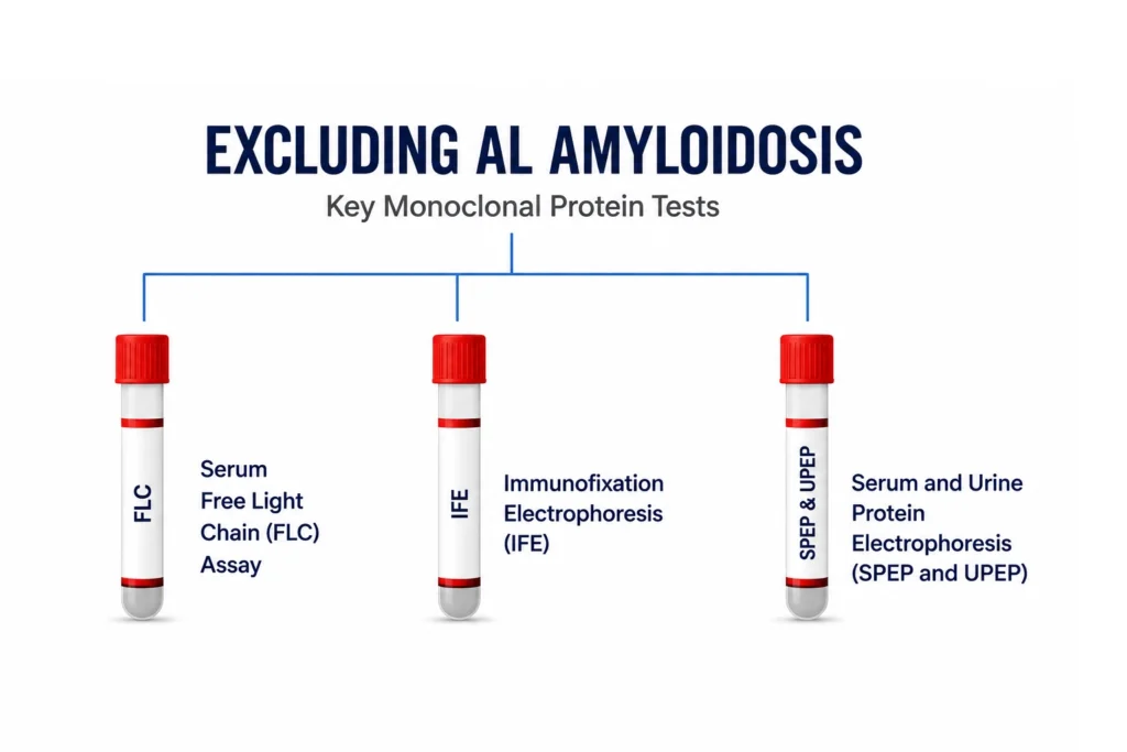
Monoclonal protein testing is a critical step in the evaluation of suspected cardiac amyloidosis. Learn how serum free light chains and serum and urine immunofixation help identify possible AL amyloidosis and guide the diagnostic pathway toward an accurate amyloid subtype diagnosis.

READ MORE

19 Aug

Cardiac Magnetic Resonance Imaging in Cardiac Amyloidosis: LGE, T1 Mapping and ECV

Cardiac magnetic resonance imaging provides detailed tissue characterization in cardiac amyloidosis. Learn how late gadolinium enhancement, native T1 mapping, extracellular volume and abnormal myocardial nulling support diagnosis and assessment.

READ MORE

19 Aug

Bone Scintigraphy Imaging of Cardiac Amyloidosis: PYP, DPD, HMDP, Perugini Grading and Diagnostic Interpretation

Bone scintigraphy is central to the non-invasive diagnosis of transthyretin cardiac amyloidosis. This article explains PYP, DPD and HMDP imaging, Perugini grading, SPECT, the H/CL ratio, monoclonal protein testing and when biopsy is still required.

READ MORE

18 Aug

Echocardiography in Cardiac Amyloidosis: Recognizing the Red Flags

Echocardiography is central to recognizing cardiac amyloidosis. Explore the key echo red flags, including increased ventricular wall thickness, restrictive filling, reduced tissue Doppler velocities, impaired global longitudinal strain, and relative apical sparing.

READ MORE