



Cardiac Magnetic Resonance Imaging in Cardiac Amyloidosis
Cardiac magnetic resonance imaging in cardiac amyloidosis provides detailed assessment of cardiac structure, ventricular function, and myocardial tissue composition. Unlike echocardiography, which primarily evaluates cardiac morphology and hemodynamics, cardiovascular magnetic resonance (CMR) can directly characterize the myocardial interstitial abnormalities associated with amyloid infiltration (1, 2).
Characteristic CMR findings include diffuse subendocardial or transmural late gadolinium enhancement (LGE), abnormal myocardial and blood-pool gadolinium kinetics, elevated native T1 values, and expansion of the extracellular volume (ECV) (1, 3).
However, CMR does not reliably determine the amyloid precursor protein by itself. Therefore, clinicians must integrate CMR findings with monoclonal protein testing, bone scintigraphy where appropriate, genetic testing, and biopsy when required to distinguish transthyretin amyloidosis (ATTR) from light-chain (AL) amyloidosis (1, 2).
Key principle: CMR is highly valuable for identifying and characterizing cardiac amyloid infiltration, but amyloid subtype determination requires integration with laboratory, nuclear imaging, genetic, and sometimes histologic findings.
What Is Cardiac Magnetic Resonance Imaging?
Cardiac magnetic resonance imaging, also called cardiovascular magnetic resonance or CMR, uses a strong magnetic field and radiofrequency signals to generate detailed images of the heart without ionizing radiation.
CMR can assess ventricular volumes, ejection fraction, wall thickness, atrial size, valve function, pericardial abnormalities, and myocardial tissue characteristics during a single examination.
Importantly, CMR does not depend on an acoustic window. Therefore, it can provide high-quality structural information when echocardiographic image quality is limited (4).
Why Is CMR Important in Cardiac Amyloidosis?
Amyloid fibrils accumulate within the myocardial extracellular space. As deposition progresses, the extracellular compartment expands and alters the distribution and washout of gadolinium contrast. CMR can detect these changes and provide information that conventional imaging cannot obtain as directly (1, 3).
As a result, CMR can:
- Raise suspicion for cardiac amyloidosis
- Characterize myocardial infiltration
- Assess ventricular morphology and function
- Quantify extracellular expansion using ECV
- Help distinguish amyloidosis from other causes of increased ventricular wall thickness
- Provide prognostic information
- Support longitudinal assessment in selected patients
What Does a CMR Examination for Cardiac Amyloidosis Include?
A comprehensive CMR examination for suspected cardiac amyloidosis usually combines several complementary techniques. Contemporary cardiomyopathy guidance recommends cine imaging, tissue characterization, T1 mapping, and late gadolinium enhancement when appropriate (4).
Important components include:
- Cine imaging for cardiac morphology and ventricular function
- Late gadolinium enhancement for myocardial infiltration patterns
- Native T1 mapping for non-contrast tissue characterization
- Post-contrast T1 mapping when ECV calculation is planned
- Extracellular volume assessment to quantify interstitial expansion
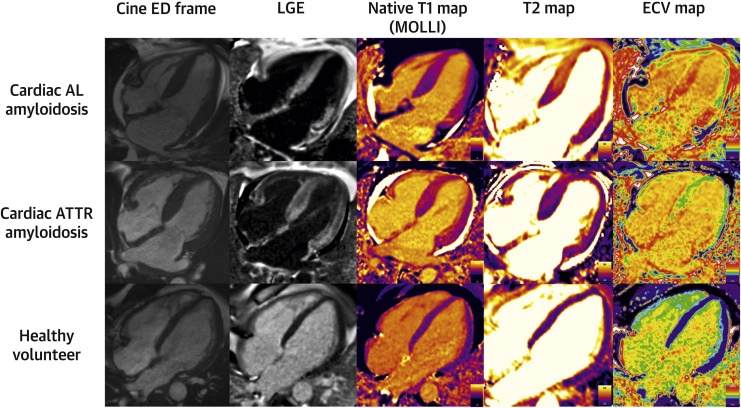
Figure 1. Multiparametric cardiovascular magnetic resonance findings in cardiac amyloidosis. Representative CMR images from a patient with cardiac light-chain (AL) amyloidosis (top row), a patient with transthyretin (ATTR) cardiac amyloidosis (middle row), and a healthy volunteer (bottom row). From left to right, the images demonstrate cine imaging at end-diastole, late gadolinium enhancement (LGE), native T1 mapping, T2 mapping, and extracellular volume (ECV) mapping. Together, these techniques demonstrate how multiparametric CMR can characterize cardiac structure, amyloid infiltration, extracellular expansion, and myocardial tissue abnormalities. Adapted from Kotecha et al., J Am Coll Cardiol. 2018;71(25):2919–2931.
Source: Kotecha T, Martinez-Naharro A, Treibel TA, et al. Myocardial edema and prognosis in amyloidosis. J Am Coll Cardiol. 2018;71(25):2919–2931. doi:10.1016/j.jacc.2018.03.536.
Cine CMR Findings in Cardiac Amyloidosis
Cine CMR provides moving images of the heart throughout the cardiac cycle. It allows accurate measurement of ventricular volumes, ejection fraction, myocardial mass, and wall thickness.
Structural findings that may occur in cardiac amyloidosis include:
- Increased left ventricular wall thickness
- Right ventricular wall thickening
- Biatrial enlargement
- Reduced ventricular cavity size in advanced restrictive disease
- Pericardial effusion
- Pleural effusion
- Progressive impairment of ventricular systolic function in advanced disease
However, these structural findings are not specific to amyloidosis. Therefore, tissue characterization is what gives CMR much of its diagnostic value.
amyloidosis (1).
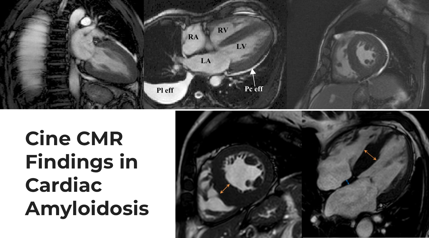
Figure 1. Cine CMR Findings in Cardiac Amyloidosis. Cine cardiac magnetic resonance imaging demonstrates characteristic structural features of cardiac amyloidosis, including increased ventricular wall thickness and altered ventricular morphology. Associated findings may include biatrial enlargement and small pleural or pericardial effusions. The images illustrate these findings across long-axis and short-axis CMR views.
Source:
Sources: Upper panels adapted from Maceira AM, Joshi J, Prasad SK, et al. Cardiovascular Magnetic Resonance in Cardiac Amyloidosis. Circulation. 2005;111(2):186–193. https://doi.org/10.1161/01.CIR.0000152819.97857.9D. Lower panels: Cardiac amyloidosis – ATTR wild type, Radiopaedia. Radiopaedia case.
Late Gadolinium Enhancement in Cardiac Amyloidosis
Late gadolinium enhancement (LGE) is one of the best-known CMR features of cardiac amyloidosis. After intravenous gadolinium administration, contrast distributes predominantly within the extracellular space. Because amyloid infiltration markedly expands this compartment, gadolinium accumulates abnormally within the myocardium (1, 3).
Characteristic LGE patterns include:
- Diffuse subendocardial enhancement
- Circumferential subendocardial enhancement
- Diffuse transmural enhancement in more advanced disease
- Patchy or segmental enhancement in some patients
- Right ventricular and atrial enhancement in more extensive disease
The ESC describes diffuse subendocardial or transmural LGE together with abnormal gadolinium kinetics as characteristic CMR findings in cardiac amyloidosis (1).
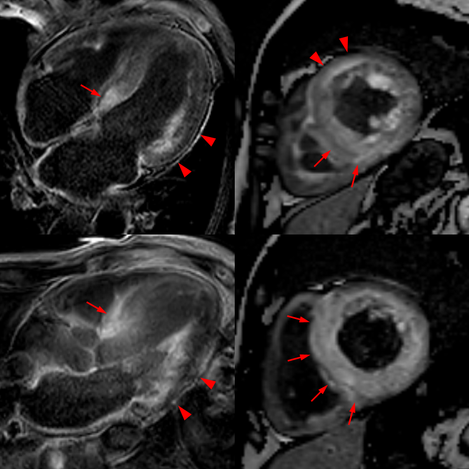
Figure 2. Late Gadolinium Enhancement in Cardiac Amyloidosis. CMR demonstrates abnormal myocardial late gadolinium enhancement (LGE) in cardiac amyloidosis. Enhancement may involve the subendocardium and can become more extensive or transmural as myocardial amyloid burden increases. The images demonstrate abnormal enhancement involving the left ventricular myocardium, with additional atrial and right ventricular involvement highlighted by the arrows and arrowheads.
Source
Source: Cardiac amyloidosis – ATTR wild type, Radiopaedia.
View the original case on Radiopaedia
Abnormal Gadolinium Kinetics and Myocardial Nulling
Cardiac amyloidosis alters not only the distribution of gadolinium but also its washout from the blood and myocardium.
During conventional inversion-recovery LGE imaging, the operator selects an inversion time that normally suppresses, or “nulls,” healthy myocardium. In cardiac amyloidosis, myocardial and blood T1 values may become unusually similar. As a result, selecting the correct myocardial nulling time can become difficult.
A characteristic abnormality is myocardial nulling before or at approximately the same time as the blood pool, rather than after the blood pool (1).
Imaging clue: Difficulty nulling the myocardium or myocardial nulling before the blood pool should raise suspicion for cardiac amyloidosis in the appropriate clinical setting.
Native T1 Mapping in Cardiac Amyloidosis
Native T1 mapping measures myocardial T1 relaxation time before gadolinium administration. Cardiac amyloid infiltration typically increases native myocardial T1 because amyloid deposition expands the extracellular compartment and alters tissue composition (3, 5).
Native T1 mapping has several important advantages. Most importantly, it does not require gadolinium contrast. Therefore, it can provide useful tissue characterization when contrast administration is undesirable or contraindicated.
However, native T1 values depend on scanner field strength, pulse sequence, vendor, and local reference ranges. Consequently, clinicians should interpret absolute T1 measurements using validated site-specific normal values rather than applying one universal cutoff (5).
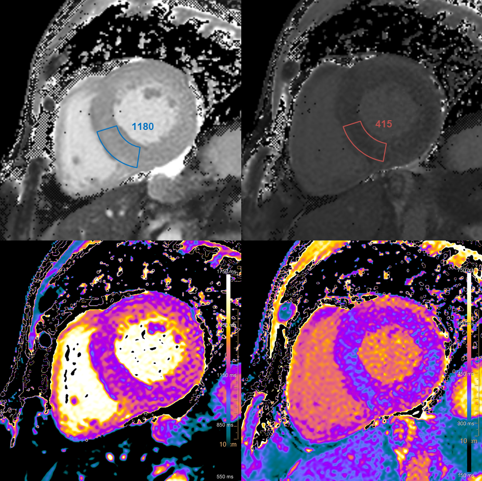
Figure 3. T1 Mapping in Transthyretin Cardiac Amyloidosis. Cardiac magnetic resonance tissue characterization in a patient with wild-type transthyretin cardiac amyloidosis. Native T1 mapping demonstrates an elevated myocardial T1 value (1180 ms), while post-contrast imaging shows a myocardial T1 value of 415 ms. T1 mapping provides quantitative assessment of myocardial tissue abnormalities associated with amyloid infiltration and contributes to extracellular volume assessment when combined with appropriate pre- and post-contrast measurements.
Source
Source: Cardiac amyloidosis – ATTR wild type, Radiopaedia.
View the original Radiopaedia case
Extracellular Volume in Cardiac Amyloidosis
Extracellular volume (ECV) is one of the most useful quantitative CMR biomarkers in cardiac amyloidosis. ECV estimates the proportion of myocardial tissue occupied by the extracellular compartment.
Because amyloid fibrils accumulate extracellularly, cardiac amyloidosis can produce marked ECV expansion. The ESC notes that an ECV of approximately 40% or greater is strongly supportive of cardiac amyloidosis, although ECV alone is not considered sufficient for diagnosis (1).
ECV can also help estimate amyloid burden and may provide useful information about disease severity and progression (4, 5).
Key distinction: LGE shows the distribution of abnormal myocardial enhancement, while T1 mapping and ECV provide quantitative information about tissue composition and extracellular expansion.
Can CMR Distinguish AL From ATTR Cardiac Amyloidosis?
CMR can strongly support the presence of cardiac amyloidosis. However, it cannot reliably determine whether the amyloid is AL or ATTR by itself.
Some differences in ventricular morphology, amyloid burden, T1 values, ECV, and enhancement patterns may occur between AL and ATTR populations. Nevertheless, there is substantial overlap between the two conditions.
Therefore, patients with suspected cardiac amyloidosis still require appropriate evaluation for a monoclonal protein and, when ATTR is suspected, bone scintigraphy and genetic testing according to the diagnostic pathway (1, 2).
CMR and the Differential Diagnosis of Increased Wall Thickness
One of the major strengths of CMR is its ability to help distinguish cardiac amyloidosis from other conditions that can produce increased ventricular wall thickness.
Hypertrophic Cardiomyopathy
Hypertrophic cardiomyopathy may produce marked myocardial thickening but generally demonstrates different patterns of hypertrophy and fibrosis. LGE is often patchy and related to hypertrophied segments rather than displaying the diffuse amyloid pattern.
Hypertensive Heart Disease
Hypertension can cause concentric left ventricular hypertrophy. However, the characteristic diffuse LGE pattern, markedly elevated T1, and pronounced ECV expansion of cardiac amyloidosis may help distinguish an infiltrative process from uncomplicated hypertensive remodeling.
Anderson-Fabry Disease
Anderson-Fabry disease can also present with increased ventricular wall thickness. Unlike cardiac amyloidosis, Fabry disease typically produces reduced native T1 values before extensive fibrosis develops, providing an important tissue-characterization clue (4, 5).
What If the Patient Has Kidney Dysfunction?
Renal impairment is common in systemic amyloidosis, particularly AL amyloidosis. Therefore, the ability to perform CMR without gadolinium can be clinically important.
Native T1 mapping and cine imaging do not require gadolinium contrast. Consequently, they can still provide valuable diagnostic information when clinicians decide not to administer gadolinium.
When contrast is being considered in patients with reduced renal function, clinicians should follow contemporary gadolinium safety guidance and select the contrast agent and imaging protocol according to individual renal function and local policy.
Can CMR Assess Disease Severity and Prognosis?
CMR provides more than diagnostic information. The extent of myocardial infiltration, LGE pattern, ventricular function, native T1, and ECV can also reflect disease severity.
In particular, extensive transmural LGE and marked extracellular volume expansion generally indicate more advanced myocardial involvement. Therefore, tissue-characterization findings may contribute to prognostic assessment when combined with biomarkers and clinical staging.
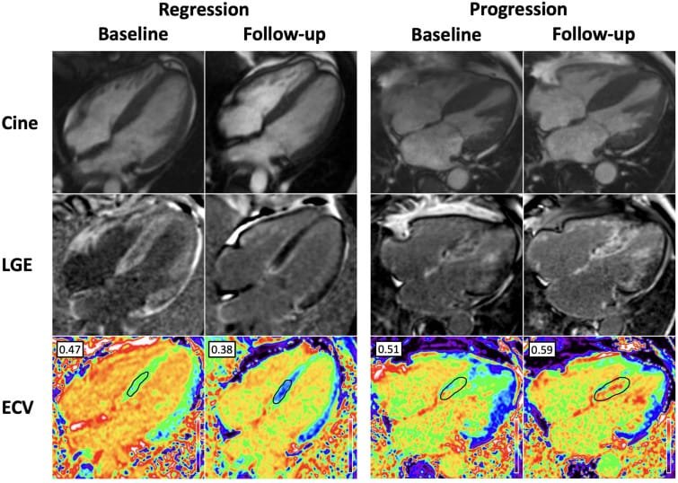
Figure 4. Regression and Progression of ATTR Cardiac Amyloidosis on CMR. Serial cardiac magnetic resonance (CMR) imaging demonstrates changes in transthyretin cardiac amyloidosis (ATTR-CA) over time. On the left, a patient receiving patisiran shows regression, with a reduction in late gadolinium enhancement (LGE) and extracellular volume (ECV) from 0.47 at baseline to 0.38 at follow-up. On the right, a patient who did not receive targeted ATTR therapy shows progression, with worsening biventricular LGE and an increase in ECV from 0.51 to 0.59. The reported normal ECV range is 0.23–0.30.
Source: Starr N, Ioannou A, Martinez-Naharro A. Monitoring cardiac amyloidosis with multimodality imaging. Revista Española de Cardiología (English Edition).
View the original article
Can CMR Monitor Cardiac Amyloidosis Over Time?
Quantitative CMR techniques, particularly ECV mapping, may help assess changes in myocardial amyloid burden over time. Consequently, CMR has potential value in monitoring disease progression and response to therapy (4).
However, serial CMR should not automatically replace established clinical assessment, biomarkers, echocardiography, or disease-specific monitoring. Its use should depend on the clinical question, local expertise, and availability.
CMR Compared With Echocardiography and Bone Scintigraphy
Echocardiography, CMR, and bone scintigraphy provide different but complementary information in suspected cardiac amyloidosis.
Echocardiography
Echocardiography is widely available and often provides the first imaging evidence of cardiac amyloidosis. It evaluates wall thickness, filling pressures, valve disease, right ventricular function, and myocardial deformation.
Cardiac Magnetic Resonance
CMR provides superior myocardial tissue characterization through LGE, native T1 mapping, and ECV assessment. Therefore, it is particularly useful when clinicians need to establish whether an infiltrative myocardial process is present.
Bone Scintigraphy
Bone scintigraphy provides a different diagnostic advantage. Grade 2 or 3 myocardial uptake with PYP, DPD, or HMDP in the absence of a monoclonal protein can establish a non-biopsy diagnosis of ATTR-CM in the appropriate clinical setting (1).
Practical distinction: Echocardiography often raises suspicion, CMR characterizes myocardial infiltration, and bone scintigraphy can establish ATTR-CM non-invasively when AL amyloidosis has been appropriately excluded.
Limitations of Cardiac Magnetic Resonance Imaging
Despite its diagnostic power, CMR has several limitations.
- Availability varies between healthcare systems.
- Image acquisition and interpretation require specialized expertise.
- Some patients cannot tolerate prolonged scanning or claustrophobic environments.
- Arrhythmias may reduce image quality in some sequences.
- Certain implanted devices may require specific MRI-compatible protocols.
- Gadolinium administration may require additional consideration in severe renal dysfunction.
- CMR alone cannot reliably distinguish ATTR from AL amyloidosis.
Cardiac Magnetic Resonance Imaging for Amyloidosis in Africa
CMR could substantially strengthen cardiac amyloidosis diagnosis across Africa by providing advanced tissue characterization in patients with unexplained ventricular wall thickening or suspected infiltrative cardiomyopathy.
However, access to cardiac-capable MRI scanners, dedicated CMR sequences, contrast agents, post-processing software, and appropriately trained specialists remains variable across the continent.
Priorities for African CMR Pathways
Potential priorities include:
- Increasing awareness of when to request CMR for suspected cardiac amyloidosis
- Developing standardized amyloidosis CMR protocols
- Expanding access to T1 mapping and ECV analysis
- Training cardiologists, radiologists, technologists, and CMR specialists
- Strengthening referral pathways between echocardiography, CMR, nuclear medicine, hematology, and pathology services
- Developing regional reference ranges for quantitative mapping where appropriate
- Improving access to amyloid typing and monoclonal protein testing alongside advanced imaging
Importantly, expanding CMR capacity should form part of a broader multimodality diagnostic pathway rather than developing as an isolated service.
A Practical CMR Checklist for Suspected Cardiac Amyloidosis
When reviewing a CMR study in suspected cardiac amyloidosis, consider:
- Is ventricular wall thickness increased?
- Are both ventricles involved?
- Is there biatrial enlargement?
- Is a pericardial or pleural effusion present?
- Is the LGE pattern diffuse subendocardial or transmural?
- Is myocardial nulling abnormal?
- Is native T1 elevated compared with local reference values?
- Is ECV markedly expanded?
- Could another cardiomyopathy better explain the findings?
- Has AL amyloidosis been evaluated with appropriate monoclonal protein testing?
- Does the patient need bone scintigraphy or biopsy to establish the amyloid subtype?
Key Takeaways
- CMR is one of the most important tissue-characterization tools in cardiac amyloidosis.
- Characteristic findings include diffuse subendocardial or transmural LGE and abnormal gadolinium kinetics.
- Native T1 is typically elevated in cardiac amyloid infiltration.
- ECV can be markedly expanded because amyloid accumulates within the extracellular compartment.
- CMR can help distinguish cardiac amyloidosis from hypertrophic cardiomyopathy, hypertensive remodeling, and Anderson-Fabry disease.
- Native T1 mapping can provide useful non-contrast tissue characterization.
- CMR may provide information about disease severity and progression.
- CMR cannot reliably distinguish AL from ATTR amyloidosis by itself.
- Therefore, laboratory testing, bone scintigraphy, genetic testing, and biopsy remain essential components of the diagnostic pathway when indicated.
Conclusion
Cardiac magnetic resonance imaging has become a central component of the multimodality assessment of cardiac amyloidosis. Through cine imaging, late gadolinium enhancement, native T1 mapping, and extracellular volume measurement, CMR can detect and quantify myocardial infiltration with a level of tissue characterization that conventional structural imaging cannot provide.
Nevertheless, CMR should not be interpreted as a stand-alone amyloid typing test. Its greatest value comes from integration with clinical findings, echocardiography, monoclonal protein testing, bone scintigraphy, genetic testing, and histology when required.
The practical principle is simple: use CMR to identify and characterize myocardial infiltration, then integrate the findings with the appropriate amyloid subtype diagnostic pathway.
References
- Garcia-Pavia P, Rapezzi C, Adler Y, et al. Diagnosis and treatment of cardiac amyloidosis: a position statement of the ESC Working Group on Myocardial and Pericardial Diseases. European Heart Journal. 2021;42(16):1554–1568. Full article
- Dorbala S, Ando Y, Bokhari S, et al. ASNC/AHA/ASE/EANM/HFSA/ISA/SCMR/SNMMI expert consensus recommendations for multimodality imaging in cardiac amyloidosis: diagnostic criteria and appropriate utilization. Journal of Nuclear Cardiology.
- Dorbala S, Cuddy S, Falk RH. How to image cardiac amyloidosis: a practical approach. Multimodality imaging consensus recommendations and standardized methods for cardiac amyloidosis.
- Arbelo E, Protonotarios A, Gimeno JR, et al. 2023 ESC Guidelines for the management of cardiomyopathies. European Heart Journal. 2023;44(37):3503–3626. Full guideline
- Moon JC, Messroghli DR, Kellman P, et al. Clinical recommendations for cardiovascular magnetic resonance mapping of T1, T2, T2* and extracellular volume: a consensus statement by the Society for Cardiovascular Magnetic Resonance endorsed by the European Association for Cardiovascular Imaging. Journal of Cardiovascular Magnetic Resonance. Full article
Medical Disclaimer
This article provides educational and informational content only and does not replace individualized medical advice, diagnosis, specialist CMR interpretation, or locally applicable imaging protocols. Clinicians should interpret CMR findings together with the patient’s clinical context, laboratory investigations, echocardiography, nuclear imaging, genetic testing, and biopsy when indicated.

[…] Cardiac MRI […]