



Monoclonal Protein Testing in Cardiac Amyloidosis
Monoclonal protein testing in cardiac amyloidosis is a critical early step in the diagnostic pathway because clinicians must rapidly identify or exclude a plasma-cell disorder that could indicate light-chain (AL) amyloidosis. AL amyloidosis and transthyretin amyloidosis (ATTR) can produce similar cardiac findings, yet their underlying biology, urgency, and treatment are fundamentally different (1, 2).
An appropriate monoclonal protein screen consists of three complementary laboratory tests: serum free light chains (sFLC), serum immunofixation electrophoresis (SIFE), and urine immunofixation electrophoresis (UIFE). When interpreted together, these tests have very high sensitivity for detecting the monoclonal protein abnormality associated with AL amyloidosis (1, 2).
Importantly, a positive monoclonal protein screen does not automatically prove AL amyloidosis. Monoclonal gammopathy of undetermined significance (MGUS) becomes increasingly common with age and can coexist with ATTR-CM. Therefore, abnormal laboratory results must be interpreted carefully and, when necessary, followed by hematologic assessment and tissue biopsy with accurate amyloid typing (1).
Key principle: In suspected cardiac amyloidosis, always evaluate for a monoclonal protein before using bone scintigraphy to establish a non-biopsy diagnosis of ATTR-CM.
Why Is Monoclonal Protein Testing Important?
The two major forms of cardiac amyloidosis are AL cardiac amyloidosis and ATTR cardiac amyloidosis. Both can cause increased ventricular wall thickness, heart failure, diastolic dysfunction, elevated cardiac biomarkers, and characteristic findings on echocardiography and cardiac magnetic resonance imaging.
However, AL amyloidosis results from a clonal plasma-cell or, less commonly, B-cell disorder that produces an amyloidogenic immunoglobulin light chain. In contrast, ATTR amyloidosis results from misfolding of transthyretin (1).
Because untreated AL amyloidosis can progress rapidly, particularly when the heart is involved, clinicians should identify a potential plasma-cell disorder early in the diagnostic process. Therefore, contemporary cardiac amyloidosis algorithms place monoclonal protein testing near the beginning of the evaluation (1, 2).
Which Tests Make Up the Monoclonal Protein Screen?
A complete monoclonal protein screen for suspected cardiac amyloidosis requires three tests:
- Serum free light-chain assay
- Serum immunofixation electrophoresis
- Urine immunofixation electrophoresis
These tests are complementary. No single component should routinely replace the others when clinicians are trying to exclude AL amyloidosis (1, 2).
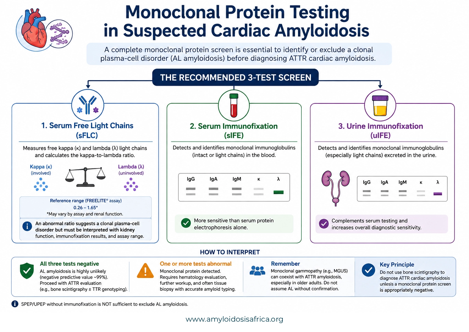
Figure 1. Monoclonal protein testing in suspected cardiac amyloidosis. A complete screen includes serum free light chains (sFLC), serum immunofixation (sIFE), and urine immunofixation (uIFE). These tests should be interpreted together when evaluating for a monoclonal gammopathy and possible AL amyloidosis. An abnormal result requires further evaluation, while negative results make AL amyloidosis highly unlikely and allow the diagnostic pathway for ATTR cardiac amyloidosis to proceed when clinically appropriate.
Serum Free Light-Chain Testing
Immunoglobulins contain two types of light chains: kappa (κ) and lambda (λ). Plasma cells normally produce small quantities of free light chains that circulate independently of intact immunoglobulins.
The serum free light-chain assay measures circulating free kappa and lambda light chains and calculates the kappa-to-lambda ratio. A clonal plasma-cell population may disproportionately produce one type of light chain, causing the ratio to become abnormal (1, 3).
Kappa and Lambda Light Chains
When the abnormal clone produces predominantly kappa light chains, serum kappa may rise disproportionately. Conversely, a lambda-producing clone may cause disproportionate elevation of serum lambda.
The light chain produced by the abnormal clone is often called the involved free light chain, whereas the other is termed the uninvolved free light chain.
The Kappa/Lambda Ratio
The free light-chain ratio is particularly useful because both kappa and lambda concentrations can rise for non-clonal reasons. Therefore, the relative balance between the two light chains provides additional information about possible clonality.
For the commonly used Freelite assay, the conventional reference range in individuals without significant kidney impairment has historically been approximately 0.26–1.65. However, reference intervals depend on the assay platform and renal function (2, 3).
Important: Do not interpret a mildly abnormal free light-chain ratio without considering kidney function, the assay used, and the serum and urine immunofixation results.
Serum Immunofixation Electrophoresis
Serum immunofixation electrophoresis (SIFE) looks for a monoclonal immunoglobulin or monoclonal light chain circulating in the blood.
Immunofixation is more sensitive than routine serum protein electrophoresis for detecting the small monoclonal protein concentrations that may occur in AL amyloidosis. Consequently, serum protein electrophoresis without immunofixation should not be used alone to exclude AL amyloidosis (1, 2).
Urine Immunofixation Electrophoresis
Urine immunofixation electrophoresis (UIFE) detects monoclonal immunoglobulin light chains excreted in the urine. Some patients with AL amyloidosis produce small quantities of monoclonal light chains that may be difficult to detect with less sensitive tests.
Therefore, urine immunofixation remains part of the recommended screening combination rather than being omitted simply because serum testing has already been performed (1, 2).
Why Are SPEP and UPEP Alone Not Enough?
Serum protein electrophoresis (SPEP) and urine protein electrophoresis (UPEP) can detect and quantify larger monoclonal protein peaks. They are valuable tests in plasma-cell disorders such as multiple myeloma.
However, the monoclonal protein burden in AL amyloidosis may be small. Consequently, conventional electrophoresis can miss abnormalities that immunofixation detects (1).
Diagnostic pitfall: A normal SPEP does not exclude AL amyloidosis. The screening pathway requires serum free light chains plus serum and urine immunofixation.
How Should Monoclonal Protein Results Be Interpreted?
Interpretation depends on the combined pattern of all three tests rather than any single result.
All Three Tests Are Negative
When serum immunofixation and urine immunofixation identify no monoclonal protein, and the serum free light-chain ratio is appropriately normal, AL amyloidosis becomes highly unlikely. The combined screening approach has a negative predictive value of approximately 99% in the diagnostic pathway described by the ACC (1).
If cardiac amyloidosis remains suspected, the patient can then proceed along the ATTR diagnostic pathway, including bone scintigraphy when appropriate.
Immunofixation Detects a Monoclonal Protein
A monoclonal band on serum or urine immunofixation indicates a monoclonal gammopathy. However, it does not by itself prove that the patient’s amyloid deposits are composed of immunoglobulin light chains.
Further evaluation is required to distinguish AL amyloidosis from MGUS, multiple myeloma, another plasma-cell disorder, or coincidental monoclonal gammopathy in a patient who actually has ATTR amyloidosis (1).
Free Light-Chain Ratio Is Abnormal
An abnormal kappa/lambda ratio can suggest monoclonal light-chain production. Nevertheless, clinicians must interpret the result alongside the absolute kappa and lambda concentrations, immunofixation studies, renal function, and the specific assay reference range.
How Does Kidney Dysfunction Affect Free Light Chains?
Kidney function is one of the most important potential confounders in serum free light-chain interpretation. The kidneys normally clear circulating free light chains. Therefore, as glomerular filtration declines, both kappa and lambda concentrations may increase even when no monoclonal plasma-cell disorder is present (2, 4).
As a result, patients with chronic kidney disease may have elevated absolute free light-chain concentrations and a mildly altered kappa/lambda ratio without true monoclonality.
Published renal reference intervals vary by assay and kidney function. For example, contemporary population data from the iStopMM study demonstrated that the expected free light-chain ratio changes progressively across eGFR categories (4).
Practical principle: A mildly abnormal free light-chain ratio in chronic kidney disease does not automatically indicate AL amyloidosis. Interpret the ratio using renal function, assay-specific reference intervals, and serum/urine immunofixation.
MGUS and ATTR Cardiac Amyloidosis
A particularly important diagnostic challenge occurs when an older patient has both suspected ATTR-CM and a monoclonal gammopathy.
Monoclonal gammopathy of undetermined significance becomes increasingly common with age. Consequently, some patients with genuine ATTR-CM also have an unrelated monoclonal protein. The ACC consensus notes that evidence of a plasma-cell dyscrasia can occur in a substantial minority of patients with ATTR-CM (1).
Therefore, a positive monoclonal screen should never automatically be equated with AL amyloidosis. Instead, clinicians must establish the actual amyloid precursor protein.
Why Monoclonal Protein Testing Must Precede ATTR Diagnosis by Bone Scintigraphy
Bone scintigraphy with PYP, DPD, or HMDP can establish ATTR-CM without biopsy in appropriately selected patients. However, this pathway is valid only when AL amyloidosis has been appropriately excluded (1, 2).
This matters because Grade 2 or Grade 3 cardiac uptake is not absolutely specific for ATTR when a monoclonal protein is present. Some patients with AL cardiac amyloidosis can show significant tracer uptake (1).
Therefore:
Grade 2–3 bone scintigraphy
+
Negative monoclonal protein screen
↓
Can support the validated non-biopsy ATTR-CM pathway
Grade 2–3 scintigraphy
+
Abnormal monoclonal protein screen
↓
Do not diagnose ATTR-CM from scintigraphy alone
+
Negative monoclonal protein screen
↓
Can support the validated non-biopsy ATTR-CM pathway
Grade 2–3 scintigraphy
+
Abnormal monoclonal protein screen
↓
Do not diagnose ATTR-CM from scintigraphy alone
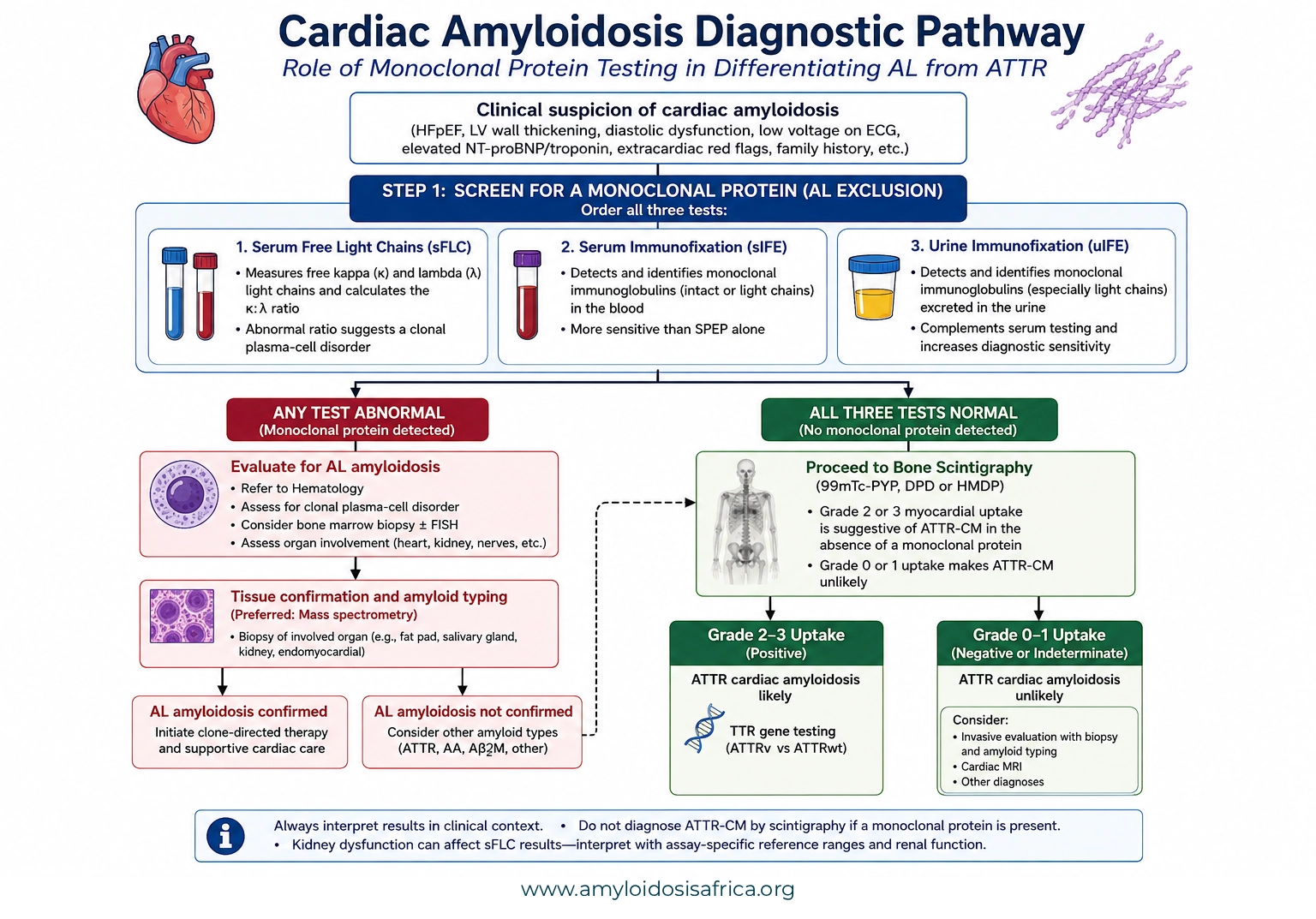
Figure 2. Cardiac amyloidosis diagnostic pathway. In patients with suspected cardiac amyloidosis, monoclonal protein screening includes serum free light chains (sFLC), serum immunofixation (sIFE), and urine immunofixation (uIFE). An abnormal result requires evaluation for a plasma-cell disorder and AL amyloidosis, with tissue confirmation and amyloid typing when appropriate. When the monoclonal protein screen is negative, bone scintigraphy can support a non-biopsy diagnosis of ATTR cardiac amyloidosis when myocardial uptake meets established diagnostic criteria.
What Happens If Monoclonal Protein Testing Is Abnormal?
An abnormal monoclonal protein screen changes the diagnostic pathway.
The patient generally requires hematology assessment to determine whether the abnormality represents:
- AL amyloidosis
- MGUS
- Multiple myeloma
- Another plasma-cell disorder
- A B-cell lymphoproliferative disorder
- A renal-function-related free light-chain abnormality rather than true monoclonality
When cardiac amyloidosis remains suspected in a patient with a monoclonal protein, tissue confirmation may be required. The biopsy specimen should not only demonstrate amyloid but should also undergo accurate amyloid typing, ideally using a validated method such as mass spectrometry where available (1).
Why Is Amyloid Typing Essential?
Finding a monoclonal protein and finding amyloid are not enough to prove that the amyloid is AL.
For example, an older patient may have ATTR amyloidosis and coincidental MGUS. If clinicians assume the monoclonal protein is responsible for the amyloid without typing the deposits, the patient could receive inappropriate therapy.
Therefore, tissue amyloid should be typed whenever the precursor protein remains uncertain. Proteomic analysis using liquid chromatography–tandem mass spectrometry provides highly specific identification of the amyloidogenic protein where available (1).
When Should AL Amyloidosis Be Strongly Suspected?
The possibility of AL amyloidosis becomes particularly important when a monoclonal protein abnormality accompanies clinical features suggestive of systemic light-chain amyloid disease.
Potential clues include:
- Unexplained proteinuria or nephrotic syndrome
- Restrictive cardiomyopathy
- Peripheral or autonomic neuropathy
- Macroglossia
- Periorbital purpura or ecchymoses
- Hepatomegaly
- Unexplained acquired factor X deficiency
- Known monoclonal gammopathy or multiple myeloma with compatible organ abnormalities
When these features occur together with abnormal monoclonal protein testing, clinicians should pursue evaluation for AL amyloidosis promptly (1).
Common Diagnostic Pitfalls
Using SPEP Alone
A normal serum protein electrophoresis result does not exclude AL amyloidosis. Serum and urine immunofixation plus serum free light chains are required for the recommended screen.
Ignoring Kidney Function
Chronic kidney disease can elevate serum free light-chain concentrations and alter the kappa/lambda ratio. Therefore, mildly abnormal free light-chain results should not automatically be interpreted as a plasma-cell clone.
Assuming Every Monoclonal Protein Means AL
MGUS is common in older adults and can coexist with ATTR-CM. Therefore, the presence of a monoclonal protein requires investigation but does not independently determine the amyloid subtype.
Interpreting a Positive PYP, DPD or HMDP Scan Without Monoclonal Testing
This is a major diagnostic error. Bone scintigraphy cannot safely distinguish ATTR-CM from AL-CM when monoclonal protein testing is abnormal or has not been performed (1, 2).
Monoclonal Protein Testing for Cardiac Amyloidosis in Africa
Reliable access to a complete monoclonal protein screen is essential for developing safe cardiac amyloidosis diagnostic pathways across Africa. Expanding bone scintigraphy without parallel access to serum free light-chain testing and immunofixation could create a risk of misclassifying AL amyloidosis as ATTR-CM.
Therefore, laboratory capacity should develop alongside imaging capacity.
Priorities for Diagnostic Pathways
- Improve access to serum free light-chain assays
- Expand serum immunofixation capacity
- Expand urine immunofixation capacity
- Avoid reliance on SPEP alone
- Develop laboratory guidance for interpreting free light chains in kidney dysfunction
- Strengthen referral pathways between cardiology, hematology, nephrology, pathology, and nuclear medicine
- Improve access to biopsy and reliable amyloid typing
- Standardize the diagnostic sequence used before ATTR-CM is diagnosed non-invasively
A Practical Monoclonal Protein Testing Checklist
Before excluding AL amyloidosis, ask:
- Were serum free kappa and lambda light chains measured?
- Was the kappa/lambda ratio calculated?
- Was serum immunofixation performed?
- Was urine immunofixation performed?
- Was kidney function considered when interpreting the free light-chain ratio?
- Was the assay-specific reference range used?
- Is there evidence of MGUS or another plasma-cell disorder?
- If any result is abnormal, has hematology been involved?
- If amyloid is demonstrated on biopsy, has the amyloid type been confirmed?
- Was ATTR-CM diagnosed by scintigraphy only after the monoclonal protein screen was appropriately negative?
Key Takeaways
- Monoclonal protein testing is essential in every patient undergoing evaluation for suspected cardiac amyloidosis.
- The recommended screen includes serum free light chains, serum immunofixation, and urine immunofixation.
- SPEP or UPEP without immunofixation is not sufficiently sensitive to exclude AL amyloidosis.
- A normal complete monoclonal protein screen makes AL amyloidosis highly unlikely.
- An abnormal monoclonal protein screen does not automatically prove AL amyloidosis.
- MGUS can coexist with ATTR cardiac amyloidosis, particularly in older patients.
- Kidney dysfunction can increase free light-chain concentrations and alter the kappa/lambda ratio.
- Bone scintigraphy should not establish ATTR-CM non-invasively when monoclonal protein testing is abnormal.
- When the amyloid subtype remains uncertain, tissue biopsy and accurate amyloid typing are essential.
Conclusion
Monoclonal protein testing is one of the most important decision points in the evaluation of suspected cardiac amyloidosis. The combination of serum free light chains, serum immunofixation, and urine immunofixation allows clinicians to identify patients who require urgent evaluation for a plasma-cell disorder and helps determine whether the validated non-biopsy ATTR-CM pathway can be used.
However, laboratory abnormalities must always be interpreted in context. Kidney dysfunction may alter serum free light chains, and MGUS may coexist with ATTR-CM. Therefore, a positive monoclonal protein screen identifies the need for further evaluation rather than establishing AL amyloidosis by itself.
The practical rule is simple: test for a monoclonal protein early, interpret all three tests together, and never use a positive bone scan to diagnose ATTR-CM until AL amyloidosis has been appropriately evaluated.
References
- Kittleson MM, Ruberg FL, Ambardekar AV, et al. 2023 ACC Expert Consensus Decision Pathway on Comprehensive Multidisciplinary Care for the Patient With Cardiac Amyloidosis. Journal of the American College of Cardiology. 2023;81(11):1076–1126. Full article
- Garcia-Pavia P, Rapezzi C, Adler Y, et al. Diagnosis and treatment of cardiac amyloidosis: a position statement of the ESC Working Group on Myocardial and Pericardial Diseases. European Heart Journal. 2021;42(16):1554–1568. Full article
- Katzmann JA, Clark RJ, Abraham RS, et al. Serum reference intervals and diagnostic ranges for free kappa and free lambda immunoglobulin light chains: relative sensitivity for detection of monoclonal light chains. Clinical Chemistry. 2002;48(9):1437–1444. PubMed
- Long TE, Indridason OS, Palsson R, et al. Defining new reference intervals for serum free light chains in individuals with chronic kidney disease: results of the iStopMM study. Blood Cancer Journal. 2022;12. PubMed
Medical Disclaimer
This article provides educational and informational content only and does not replace individualized medical advice, laboratory interpretation, hematology consultation, or locally applicable diagnostic guidelines. Free light-chain results should be interpreted using the assay-specific reference range, kidney function, immunofixation findings, and the patient’s overall clinical context.

[…] Monoclonal Protein Testing […]