



ATTR vs AL Amyloidosis: Key Differences in Causes, Red Flags, Diagnosis and Treatment
AL amyloidosis and transthyretin amyloidosis (ATTR) are two of the most important forms of systemic amyloidosis, and both can involve the heart. Although they may produce similar clinical findings, they arise from completely different precursor proteins and require fundamentally different treatment strategies. (1, 2)
Correctly distinguishing AL from ATTR amyloidosis is therefore one of the most important steps in the diagnostic evaluation of a patient with suspected cardiac amyloidosis. Misclassification can delay appropriate disease-specific treatment. (2, 3)
What Is AL Amyloidosis?
Immunoglobulin light-chain amyloidosis, or AL amyloidosis, is caused by abnormal immunoglobulin light chains produced by a clonal population of plasma cells or, less commonly, another B-cell clone. These light chains or their fragments misfold and form amyloid fibrils that deposit within tissues and organs. (4)
AL amyloidosis is therefore fundamentally a hematologic disorder. It can involve multiple organs, including the heart, kidneys, peripheral and autonomic nervous systems, gastrointestinal tract, liver and soft tissues. (4)
Cardiac involvement is particularly important because it is a major determinant of prognosis. AL amyloidosis can progress rapidly, making timely recognition and hematologic assessment essential. (4)
What Is ATTR Amyloidosis?
Transthyretin amyloidosis (ATTR) is caused by misfolding and aggregation of transthyretin, a transport protein produced predominantly by the liver. ATTR occurs in two major forms. (5)
- Wild-type ATTR amyloidosis (ATTRwt) – occurs without a pathogenic TTR gene variant and predominantly affects older adults.
- Variant/hereditary ATTR amyloidosis (ATTRv) – results from a pathogenic or likely pathogenic variant in the TTR gene.
ATTR may primarily affect the heart, peripheral nerves, autonomic nervous system or a combination of these systems, depending on the form and specific genetic variant. (5)
Core distinction
AL amyloidosis: abnormal immunoglobulin light chains produced by a plasma cell or other B-cell clone.
ATTR amyloidosis: misfolded transthyretin, either wild-type or genetically variant.
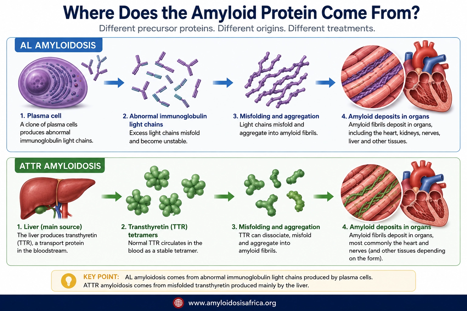
AL vs ATTR Amyloidosis: Key Differences at a Glance
How Do Their Clinical Presentations Differ?
There is substantial overlap between AL and ATTR amyloidosis, particularly when the heart is involved. Both can cause increased ventricular wall thickness, restrictive cardiac physiology, heart failure, atrial arrhythmias and conduction abnormalities. (2, 3)
The extracardiac phenotype can nevertheless provide important clues.
Features That May Raise Suspicion for AL Amyloidosis
- Proteinuria or nephrotic syndrome
- Peripheral neuropathy
- Autonomic dysfunction
- Macroglossia
- Periorbital purpura
- Unexplained hepatomegaly
- Multisystem disease occurring with a monoclonal gammopathy
Macroglossia and periorbital purpura are classically associated with AL amyloidosis and may be particularly suggestive when present, although neither occurs in every patient. (4)
Features That May Raise Suspicion for ATTR Amyloidosis
- Bilateral carpal tunnel syndrome
- Lumbar spinal stenosis
- Spontaneous distal biceps tendon rupture
- Cardiomyopathy in an older adult
- Peripheral neuropathy
- Autonomic dysfunction
- Family history of amyloidosis, neuropathy or cardiomyopathy
Musculoskeletal manifestations such as carpal tunnel syndrome and lumbar spinal stenosis may precede recognition of ATTR cardiomyopathy by several years. (6)
Important: These red flags are suggestive, not diagnostic. AL and ATTR can overlap clinically, and the amyloid type should never be assigned solely from symptoms or imaging appearance.
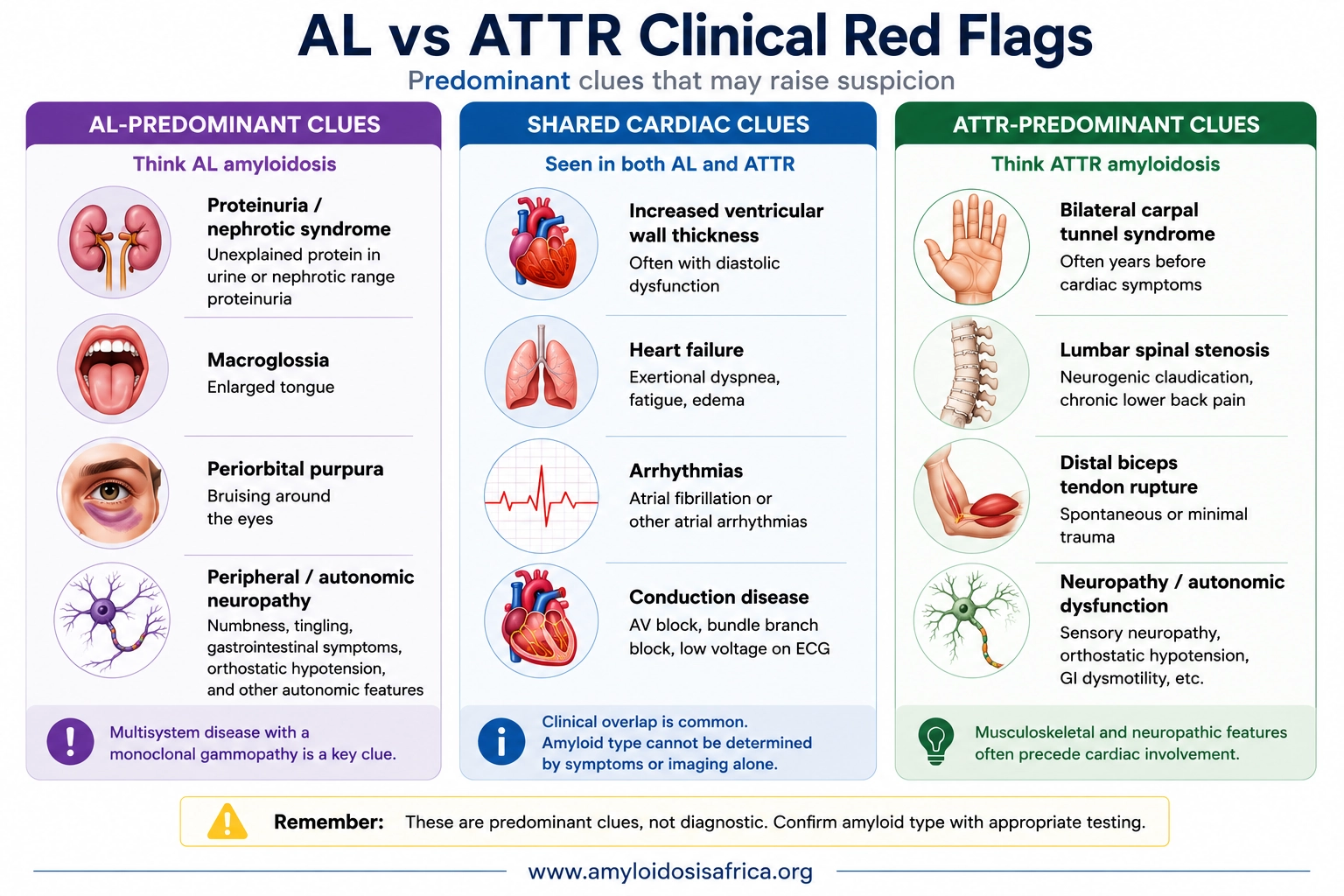
Can ECG Distinguish AL From ATTR?
No. ECG abnormalities can increase suspicion for cardiac amyloidosis but generally cannot reliably distinguish AL from ATTR. Potential findings include low QRS voltage, pseudoinfarction patterns, atrial fibrillation and conduction abnormalities. (2)
Importantly, low voltage is not present in all patients with cardiac amyloidosis and should not be considered mandatory for diagnosis.
Can Echocardiography Distinguish AL From ATTR?
Echocardiography may raise strong suspicion for cardiac amyloidosis but usually cannot determine the amyloid precursor protein by itself. Common findings include increased ventricular wall thickness, diastolic dysfunction, biatrial enlargement and reduced longitudinal strain. (3)
Relative apical sparing of longitudinal strain can support suspicion for cardiac amyloidosis but should not be used as a standalone test to distinguish AL from ATTR.
Can Cardiac MRI Distinguish AL From ATTR?
Cardiac magnetic resonance can strongly support the presence of cardiac amyloidosis through findings such as diffuse late gadolinium enhancement, abnormal gadolinium kinetics, elevated native T1 and increased extracellular volume. (3)
Although some imaging characteristics may differ statistically between AL and ATTR populations, CMR alone generally cannot provide definitive amyloid typing. Further laboratory, scintigraphic or tissue evaluation is required.
The Critical Diagnostic Question: Is a Monoclonal Protein Present?
When cardiac amyloidosis is suspected, assessment for a monoclonal immunoglobulin is a critical early step because AL amyloidosis requires urgent consideration. Recommended testing includes (2, 3):
- Serum free light-chain assay
- Serum immunofixation electrophoresis
- Urine immunofixation electrophoresis
Serum protein electrophoresis alone is not an adequate test to exclude AL amyloidosis. A complete monoclonal protein assessment should include serum free light chains plus serum and urine immunofixation.
Does a Monoclonal Protein Automatically Mean AL Amyloidosis?
No. A monoclonal gammopathy can coexist with ATTR amyloidosis, particularly in older adults. Therefore, detecting a monoclonal protein does not by itself establish that the amyloid deposits are AL. (2)
When a monoclonal protein is present and cardiac amyloidosis is suspected, additional specialist evaluation and, in many cases, tissue biopsy with definitive amyloid typing may be necessary.
How Bone Scintigraphy Helps Identify ATTR-CM
Bone-avid tracer scintigraphy with validated tracers such as 99mTc-PYP, 99mTc-DPD or 99mTc-HMDP is highly valuable in the diagnostic evaluation of ATTR cardiac amyloidosis. (7)
The landmark multicentre study by Gillmore and colleagues demonstrated that, in an appropriate clinical setting, grade 2 or 3 cardiac uptake together with the absence of a monoclonal protein can establish a non-biopsy diagnosis of ATTR cardiac amyloidosis with very high specificity. (7)
A positive bone scintigraphy scan alone does not prove ATTR-CM. Some patients with AL amyloidosis can also demonstrate cardiac tracer uptake. Monoclonal protein testing is therefore an essential part of interpreting the scan.
When Is a Biopsy Needed?
Tissue biopsy becomes particularly important when the non-biopsy ATTR criteria are not fulfilled, when a monoclonal gammopathy is present, when imaging findings are discordant or when the amyloid type remains uncertain. (2, 3)
Amyloid deposits are traditionally identified using Congo red staining. However, simply proving that amyloid is present is insufficient. The deposited protein must also be accurately typed.
Mass spectrometry-based proteomic analysis is an important reference method for amyloid typing where available. (1)
A Practical AL vs ATTR Diagnostic Pathway
Patient with suspected cardiac amyloidosis
↓
Perform monoclonal protein assessment
Serum free light chains + serum immunofixation + urine immunofixation
↓
If ATTR-CM remains suspected
Perform validated bone-avid tracer scintigraphy
↓
Grade 2/3 uptake + no monoclonal protein
ATTR-CM may be diagnosed noninvasively in the appropriate clinical context
↓
Monoclonal protein present or non-biopsy criteria not fulfilled
Further specialist assessment ± tissue biopsy and amyloid typing
↓
If ATTR is confirmed
Perform TTR genetic testing to distinguish ATTRv from ATTRwt
This is a simplified educational pathway and should be interpreted alongside detailed guidelines and the individual patient’s clinical context. (2, 3, 7)
How Does Treatment Differ?
The treatment of AL and ATTR amyloidosis is fundamentally different because the precursor proteins arise from different biological processes.
Treatment of AL Amyloidosis
Treatment of AL amyloidosis targets the abnormal plasma-cell or B-cell clone responsible for producing the amyloidogenic immunoglobulin light chains. The therapeutic objective is rapid and deep suppression of light-chain production. (4)
Modern therapy commonly uses a daratumumab-based combination including bortezomib, cyclophosphamide and dexamethasone in appropriate newly diagnosed patients. Autologous stem-cell transplantation remains an option for carefully selected patients. (4)
Treatment of ATTR Amyloidosis
ATTR treatment targets the transthyretin disease pathway rather than a plasma-cell clone. Strategies include stabilizing circulating transthyretin, reducing hepatic TTR production and managing organ-specific complications. (5)
Treatment selection depends on whether the patient has cardiomyopathy, polyneuropathy or a mixed phenotype, as well as disease severity, regulatory approval, availability and individual clinical factors.
Why Is Correct Amyloid Typing So Important?
AL and ATTR amyloidosis may look similar clinically, especially when both present as cardiac amyloidosis. However, treatment for one does not treat the underlying cause of the other.
Accurate amyloid typing prevents inappropriate therapy and allows disease-specific treatment to begin as early as possible. For this reason, major cardiac amyloidosis guidance emphasizes defining the amyloid precursor protein rather than stopping once cardiac amyloid involvement is suspected. (2, 3)
ATTR and AL Amyloidosis in Africa
The burden and distribution of both AL and ATTR amyloidosis across African countries remain incompletely characterized. Limited epidemiological data, variable access to specialized diagnostics and constrained availability of amyloid typing, nuclear scintigraphy and genetic testing can make accurate subtype diagnosis challenging in some settings. (8)
Hereditary ATTR is particularly relevant to African populations because the TTR p.Val142Ile variant, historically referred to as V122I, is associated with West African ancestry. A population genetic analysis demonstrated substantial variation in allele frequency across African populations, with comparatively higher frequencies in parts of West Africa. (9)
The presence of a TTR variant should not be equated with clinically manifest amyloidosis because penetrance is incomplete and clinical expression varies. Likewise, genetic carrier frequency should not be used as a substitute for direct epidemiological studies of ATTR-CM in African populations.
Strengthening access to appropriate monoclonal protein testing, cardiac imaging, amyloid typing, scintigraphy and genetic testing will be important for improving accurate differentiation of AL and ATTR amyloidosis across African healthcare systems.
Key Takeaways
- AL and ATTR amyloidosis are different diseases caused by different precursor proteins.
- AL amyloidosis results from abnormal immunoglobulin light chains produced by a plasma-cell or other B-cell clone.
- ATTR amyloidosis results from transthyretin misfolding and occurs as ATTRwt or hereditary ATTRv.
- Both AL and ATTR can cause cardiac amyloidosis and may appear similar on ECG, echocardiography and CMR.
- Proteinuria, macroglossia and periorbital purpura may raise suspicion for AL amyloidosis.
- Bilateral carpal tunnel syndrome, lumbar spinal stenosis and distal biceps tendon rupture may raise suspicion for ATTR amyloidosis.
- Clinical red flags alone cannot establish amyloid type.
- A complete monoclonal protein screen includes serum free light chains, serum immunofixation and urine immunofixation.
- A monoclonal gammopathy does not automatically prove AL amyloidosis because monoclonal gammopathy can coexist with ATTR.
- A positive bone scintigraphy scan alone does not establish ATTR-CM.
- Grade 2 or 3 cardiac uptake on validated bone scintigraphy together with the absence of a monoclonal protein can establish ATTR-CM noninvasively in the appropriate clinical context.
- Tissue biopsy and definitive amyloid typing may be required when the diagnostic pathway is inconclusive or a monoclonal gammopathy is present.
- Once ATTR is confirmed, TTR genetic testing distinguishes ATTRv from ATTRwt.
- Correct amyloid typing is essential because AL and ATTR require fundamentally different disease-specific treatments.
References
- Buxbaum JN, Eisenberg DS, Fändrich M, McPhail ED, Merlini G, Saraiva MJM, et al. Amyloid nomenclature 2024: update, novel proteins, and recommendations by the International Society of Amyloidosis (ISA) Nomenclature Committee. Amyloid. 2024;31(4):249-256. doi:10.1080/13506129.2024.2405948.
- Kittleson MM, Ruberg FL, Ambardekar AV, Brannagan TH 3rd, Cheng RK, Clarke JO, et al. 2023 ACC Expert Consensus Decision Pathway on comprehensive multidisciplinary care for the patient with cardiac amyloidosis: a report of the American College of Cardiology Solution Set Oversight Committee. J Am Coll Cardiol. 2023;81(11):1076-1126. doi:10.1016/j.jacc.2022.11.022.
- Garcia-Pavia P, Rapezzi C, Adler Y, Arad M, Basso C, Brucato A, et al. Diagnosis and treatment of cardiac amyloidosis: a position statement of the ESC Working Group on Myocardial and Pericardial Diseases. Eur Heart J. 2021;42(16):1554-1568. doi:10.1093/eurheartj/ehab072.
- Gertz MA. Immunoglobulin light chain amyloidosis: 2024 update on diagnosis, prognosis, and treatment. Am J Hematol. 2024;99(2):309-324. doi:10.1002/ajh.27177.
- Ruberg FL, Maurer MS. Cardiac amyloidosis due to transthyretin protein: a review. JAMA. 2024;331(9):778-791. doi:10.1001/jama.2024.0442.
- Ruberg FL, Grogan M, Hanna M, Kelly JW, Maurer MS. Transthyretin amyloid cardiomyopathy: JACC state-of-the-art review. J Am Coll Cardiol. 2019;73(22):2872-2891. doi:10.1016/j.jacc.2019.04.003.
- Gillmore JD, Maurer MS, Falk RH, Merlini G, Damy T, Dispenzieri A, et al. Nonbiopsy diagnosis of cardiac transthyretin amyloidosis. Circulation. 2016;133(24):2404-2412. doi:10.1161/CIRCULATIONAHA.116.021612.
- Madu EC, Mezue K. Uneven burden of cardiac amyloidosis in people of African descent: global imbalance in resources and access. BMC Glob Public Health. 2023;1:15. doi:10.1186/s44263-023-00016-3.
- Jacobson DR, Alexander AA, Tagoe C, Garvey WT, Williams SM, Tishkoff S, et al. The prevalence and distribution of the amyloidogenic transthyretin (TTR) V122I allele in Africa. Mol Genet Genomic Med. 2016;4(5):548-556. doi:10.1002/mgg3.231.
Medical Disclaimer
This article is intended for educational and informational purposes and is not a substitute for individualized medical advice, diagnosis or treatment. The investigation and management of suspected amyloidosis should be guided by appropriately qualified healthcare professionals, specialist assessment and applicable local clinical guidance.

Leave a Reply