



How Is Cardiac Amyloidosis Diagnosed? A Step-by-Step Diagnostic Approach
Diagnosing cardiac amyloidosis requires more than identifying a thickened heart on echocardiography. The diagnostic process must establish whether cardiac amyloidosis is present and, critically, determine the underlying amyloid type because the diagnostic pathways and treatments for transthyretin amyloidosis (ATTR) and immunoglobulin light-chain amyloidosis (AL) are fundamentally different (1, 2).
Modern diagnostic algorithms combine clinical suspicion, electrocardiography, echocardiography, cardiac magnetic resonance imaging, monoclonal protein testing, bone-avid tracer scintigraphy and, when necessary, tissue biopsy and definitive amyloid typing (1, 2).
Key principle: Diagnosing cardiac amyloidosis involves two questions: Is amyloid affecting the heart? and what type of amyloid is responsible?
Step 1: Recognize When Cardiac Amyloidosis Should Be Suspected
The diagnostic pathway begins with clinical suspicion. Cardiac amyloidosis should be considered when cardiac findings occur together with compatible systemic or extracardiac features (1, 2).
Potential clues include:
- Unexplained increased left ventricular wall thickness
- Heart failure, particularly with preserved or mildly reduced ejection fraction
- Atrial fibrillation
- Conduction disease or unexplained pacemaker requirement
- Bilateral carpal tunnel syndrome
- Lumbar spinal stenosis
- Spontaneous distal biceps tendon rupture
- Peripheral or autonomic neuropathy
- Proteinuria or nephrotic syndrome
- Macroglossia or periorbital purpura, particularly suggestive of AL amyloidosis
No individual red flag is sufficient to diagnose cardiac amyloidosis. Suspicion becomes stronger when multiple compatible findings occur together.
Step 2: Electrocardiography
An electrocardiogram (ECG) is part of the initial cardiac assessment. Potential abnormalities include conduction disease, atrial fibrillation, pseudoinfarction patterns and reduced QRS voltage.
The classic teaching of low ECG voltage despite increased ventricular wall thickness can be an important clue, but low voltage is not present in every patient. Its absence therefore does not exclude cardiac amyloidosis (1).
Important: A normal or non-specific ECG does not rule out cardiac amyloidosis.
Step 3: Echocardiography
Echocardiography is one of the most important initial imaging investigations when cardiac amyloidosis is suspected.
Potential echocardiographic findings include:
- Increased left ventricular wall thickness
- Right ventricular wall thickening
- Biatrial enlargement
- Diastolic dysfunction
- Reduced longitudinal systolic function despite preserved ejection fraction
- Valve thickening
- Small pericardial effusion
Global Longitudinal Strain and Apical Sparing
Speckle-tracking echocardiography may demonstrate reduced global longitudinal strain with relative preservation of apical longitudinal strain, producing the characteristic relative apical sparing pattern.
This pattern can strengthen suspicion for cardiac amyloidosis, but it is not sufficiently specific to establish the diagnosis by itself (3).
Step 4: Cardiac Magnetic Resonance Imaging
Cardiac magnetic resonance (CMR) provides detailed structural and tissue characterization and can strengthen the diagnosis when cardiac amyloidosis is suspected.
Features that may support cardiac amyloidosis include:
- Diffuse subendocardial or transmural late gadolinium enhancement
- Abnormal myocardial and blood-pool gadolinium kinetics
- Elevated native T1 values
- Expansion of extracellular volume
CMR is particularly valuable for identifying an infiltrative cardiomyopathy and assessing disease burden. However, CMR generally cannot reliably determine whether the amyloid precursor protein is AL or ATTR. Amyloid typing therefore requires the appropriate laboratory, scintigraphic or histological pathway (2).
Step 5: Screen for a Monoclonal Protein
This is one of the most important steps in the entire diagnostic pathway. When cardiac amyloidosis is suspected, clinicians must assess for evidence of a monoclonal immunoglobulin that could indicate a plasma cell disorder and possible AL amyloidosis (1).
Complete monoclonal protein screen
- Serum free light-chain assay
- Serum immunofixation electrophoresis
- Urine immunofixation electrophoresis
When serum and urine immunofixation show no monoclonal protein and the serum free light-chain ratio is normal, AL amyloidosis is essentially excluded with very high negative predictive value (1).
Why SPEP Alone Is Not Enough
A common diagnostic mistake is relying on standard serum protein electrophoresis (SPEP) or urine protein electrophoresis (UPEP) alone to exclude a monoclonal protein.
Do not use SPEP/UPEP alone to exclude AL amyloidosis.
The recommended monoclonal protein screen includes serum free light chains plus serum and urine immunofixation (1).
The recommended monoclonal protein screen includes serum free light chains plus serum and urine immunofixation (1).
What If the Monoclonal Protein Screen Is Abnormal?
An abnormal monoclonal protein screen does not automatically establish AL amyloidosis. Monoclonal gammopathy of undetermined significance (MGUS), kidney dysfunction and other plasma cell disorders may complicate interpretation.
However, if a monoclonal protein is detected or the serum free light-chain ratio is abnormal, AL amyloidosis must be evaluated promptly, usually with haematology involvement and tissue confirmation with definitive amyloid typing (1, 2).
Step 6: Bone-Avid Tracer Scintigraphy for Suspected ATTR-CM
When the monoclonal protein screen is negative and ATTR cardiomyopathy remains suspected, bone-avid tracer scintigraphy can provide a non-invasive diagnosis in appropriately selected patients (4).
Validated tracers include:
- 99mTc-PYP – technetium-99m pyrophosphate
- 99mTc-DPD – technetium-99m DPD
- 99mTc-HMDP – technetium-99m hydroxymethylene diphosphonate
Understanding Visual Scintigraphy Grades
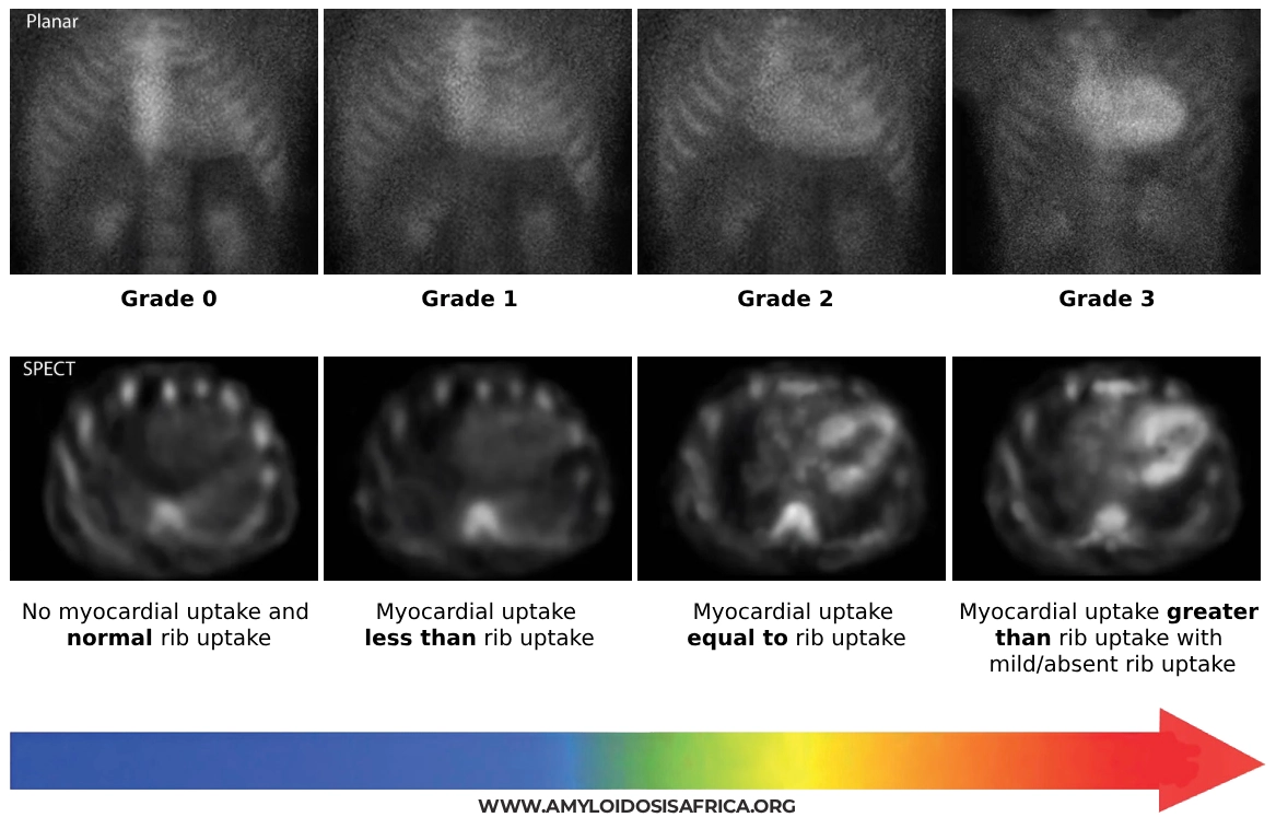
Grade 2 or 3 myocardial uptake can support a non-biopsy diagnosis of ATTR-CM only when a monoclonal protein has been appropriately excluded and the patient has a compatible cardiac phenotype (4).
A positive scintigraphy scan alone does not diagnose ATTR-CM. AL cardiac amyloidosis can also demonstrate tracer uptake. The result must therefore be interpreted together with the monoclonal protein screen.
Why SPECT or SPECT/CT Matters
Planar scintigraphy can sometimes mistake residual radiotracer within the cardiac blood pool for true myocardial uptake.
Single-photon emission computed tomography (SPECT), with or without CT, allows three-dimensional assessment and helps confirm that tracer activity is located within the myocardium rather than the ventricular blood pool (1).
Step 7: When Is Biopsy Required?
Although ATTR-CM can sometimes be diagnosed without biopsy, histological confirmation remains essential in several clinical scenarios (1, 2).
Biopsy should be considered when:
- A monoclonal protein is present and cardiac amyloidosis is suspected
- Scintigraphy is negative or equivocal despite high clinical suspicion
- Scintigraphy and laboratory findings are discordant
- The non-biopsy diagnostic criteria for ATTR-CM are not fulfilled
- Bone-avid tracer scintigraphy is unavailable and definitive diagnosis is required
Congo Red Staining
Histological diagnosis is based on demonstrating amyloid deposits in tissue. Congo red staining classically produces apple-green birefringence when examined under polarized light.
Potential biopsy sites include abdominal fat, bone marrow or a clinically affected organ. The sensitivity of extracardiac biopsy varies according to the amyloid type, and a negative fat-pad biopsy does not exclude cardiac amyloidosis (1).
Amyloid Typing Is Essential
Demonstrating amyloid in tissue is only part of the diagnosis. The amyloid precursor protein must also be correctly identified because treatment differs substantially between AL, ATTR and other amyloid types.
Proteomic analysis using laser microdissection followed by mass spectrometry is a highly accurate method for definitive amyloid typing and is particularly valuable when immunohistochemical results are uncertain (5).
Step 8: Genetic Testing After ATTR Is Confirmed
Once ATTR amyloidosis has been established, TTR gene sequencing should be performed to distinguish hereditary ATTR (ATTRv) from wild-type ATTR (ATTRwt) (1, 2).
Genetic testing remains important even in older patients who appear clinically likely to have wild-type ATTR because identification of a pathogenic TTR variant has implications for the patient and potentially for biological relatives.
Genetic testing comes after ATTR is established. It distinguishes ATTRv from ATTRwt; it should not be used by itself to prove that cardiac amyloid deposition is present.
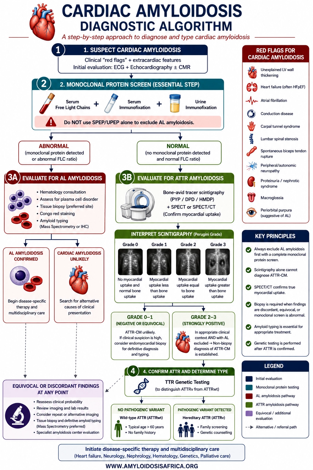
The Cardiac Amyloidosis Diagnostic Algorithm
1. Suspect cardiac amyloidosis
Clinical red flags + ECG + echocardiography ± CMR
↓
2. Perform a complete monoclonal protein screen
Serum free light chains + serum immunofixation + urine immunofixation
↓
3A. Monoclonal screen abnormal
Evaluate urgently for AL amyloidosis → haematology assessment → tissue biopsy and definitive amyloid typing when indicated
OR
3B. Monoclonal screen negative
If ATTR-CM remains suspected → PYP / DPD / HMDP scintigraphy with SPECT or SPECT/CT
↓
4. Grade 2 or 3 myocardial uptake
In the appropriate clinical context and with AL excluded → non-biopsy diagnosis of ATTR-CM
↓
5. ATTR confirmed
TTR genetic testing → ATTRwt or ATTRv
↓
If findings are equivocal or discordant
Specialist evaluation ± tissue biopsy and definitive amyloid typing
Common Diagnostic Pitfalls
Pitfall 1: Waiting for low ECG voltage
Low voltage is not present in every patient with cardiac amyloidosis.
Pitfall 2: Assuming apical sparing proves amyloidosis
Relative apical sparing is a useful clue but is not diagnostic by itself.
Pitfall 3: Using SPEP alone to exclude AL
A complete monoclonal protein screen requires serum free light chains plus serum and urine immunofixation.
Pitfall 4: Diagnosing ATTR from a positive PYP/DPD/HMDP scan alone
AL amyloidosis must be appropriately excluded before scintigraphy can establish a non-biopsy ATTR-CM diagnosis.
Pitfall 5: Relying on planar scintigraphy alone
Blood-pool activity can mimic myocardial uptake. SPECT or SPECT/CT helps confirm true myocardial localization.
Pitfall 6: Assuming a negative fat-pad biopsy excludes cardiac amyloidosis
The sensitivity of extracardiac biopsy varies by amyloid type. Further investigation may be required when clinical suspicion remains high.
Pitfall 7: Failing to type amyloid found on biopsy
Confirmation of amyloid deposition should be followed by accurate identification of the amyloid precursor protein.
Diagnosing Cardiac Amyloidosis in Africa
The principles of cardiac amyloidosis diagnosis are applicable across healthcare settings, but implementation depends on local access to laboratory testing, advanced cardiac imaging, nuclear medicine, pathology, mass spectrometry and genetic testing.
Important priorities for strengthening diagnostic capacity across Africa include:
- Increasing awareness of cardiac and extracardiac red flags
- Expanding access to high-quality echocardiography and strain imaging
- Ensuring access to serum free light-chain testing and serum and urine immunofixation
- Expanding appropriately performed bone-avid tracer scintigraphy where feasible
- Strengthening pathology and amyloid-typing capacity
- Improving access to TTR genetic testing and genetic counselling
- Developing clear referral pathways to specialist amyloidosis centres and multidisciplinary teams
Key Takeaways
- Cardiac amyloidosis diagnosis begins with clinical suspicion.
- ECG and echocardiography provide important clues but cannot establish the amyloid type.
- CMR provides valuable tissue characterization but generally cannot reliably distinguish AL from ATTR.
- A complete monoclonal protein screen requires serum free light chains, serum immunofixation and urine immunofixation.
- SPEP/UPEP alone should not be used to exclude AL amyloidosis.
- Grade 2 or 3 myocardial uptake on validated bone scintigraphy can establish ATTR-CM noninvasively when AL has been appropriately excluded and the clinical phenotype is compatible.
- SPECT or SPECT/CT helps distinguish true myocardial tracer uptake from blood-pool activity.
- Biopsy is required when non-invasive criteria are not fulfilled or findings are discordant.
- Amyloid identified on biopsy should be accurately typed.
- After ATTR is confirmed, TTR genetic testing distinguishes hereditary ATTR from wild-type ATTR.
References
- Kittleson MM, Ruberg FL, Ambardekar AV, Brannagan TH 3rd, Cheng RK, Clarke JO, et al. 2023 ACC Expert Consensus Decision Pathway on comprehensive multidisciplinary care for the patient with cardiac amyloidosis. J Am Coll Cardiol. 2023;81(11):1076-1126. doi:10.1016/j.jacc.2022.11.022.
- Garcia-Pavia P, Rapezzi C, Adler Y, Arad M, Basso C, Brucato A, et al. Diagnosis and treatment of cardiac amyloidosis: a position statement of the ESC Working Group on Myocardial and Pericardial Diseases. Eur Heart J. 2021;42(16):1554-1568. doi:10.1093/eurheartj/ehab072.
- Phelan D, Collier P, Thavendiranathan P, Popović ZB, Hanna M, Plana JC, et al. Relative apical sparing of longitudinal strain using two-dimensional speckle-tracking echocardiography is both sensitive and specific for the diagnosis of cardiac amyloidosis. Heart. 2012;98(19):1442-1448. doi:10.1136/heartjnl-2012-302353.
- Gillmore JD, Maurer MS, Falk RH, Merlini G, Damy T, Dispenzieri A, et al. Nonbiopsy diagnosis of cardiac transthyretin amyloidosis. Circulation. 2016;133(24):2404-2412. doi:10.1161/CIRCULATIONAHA.116.021612.
- Vrana JA, Gamez JD, Madden BJ, Theis JD, Bergen HR 3rd, Dogan A. Classification of amyloidosis by laser microdissection and mass spectrometry-based proteomic analysis in clinical biopsy specimens. Blood. 2009;114(24):4957-4959. doi:10.1182/blood-2009-07-230722.
Medical Disclaimer
This article is intended for educational and informational purposes and is not a substitute for individualized medical advice, diagnosis or treatment. Diagnostic decisions should be made by appropriately qualified healthcare professionals based on the individual patient’s clinical circumstances, locally applicable guidance and available diagnostic resources.

Leave a Reply