



AL Amyloidosis: Causes, Symptoms, Diagnosis and Treatment
AL amyloidosis is a systemic disease caused by abnormal immunoglobulin light chains produced by a clonal population of plasma cells or, less commonly, another B-cell disorder. These unstable light chains can misfold, form amyloid fibrils and accumulate in tissues. As a result, AL amyloidosis can damage the heart, kidneys, peripheral nerves, gastrointestinal tract, liver and other organs (1, 2).
Importantly, cardiac involvement is one of the major determinants of prognosis. Unlike transthyretin amyloidosis, which results from misfolded transthyretin, AL amyloidosis requires treatment directed at the abnormal plasma-cell or B-cell clone (3).
Key principle: Clinicians should recognize suspected AL amyloidosis promptly because continued production of toxic amyloidogenic light chains can cause progressive and sometimes rapidly worsening organ dysfunction.
What Is AL Amyloidosis?
The abbreviation AL refers to amyloid derived from immunoglobulin light chains. Normally, plasma cells produce antibodies that contain heavy and light chains as part of the immune response.
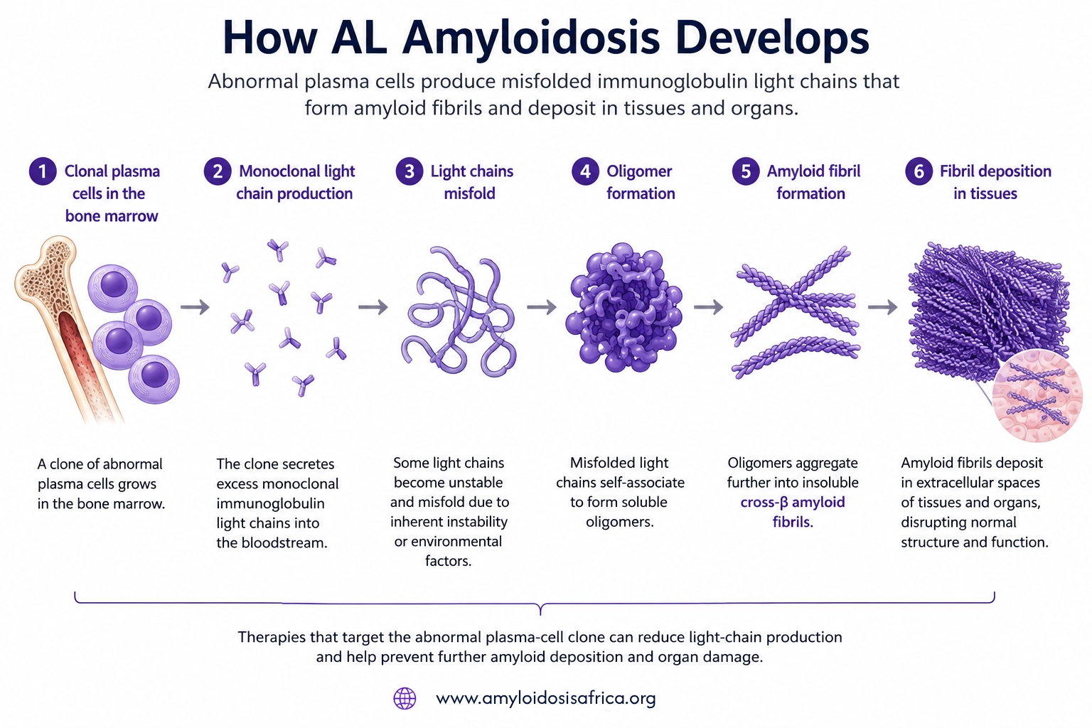
In AL amyloidosis, however, an abnormal clone of plasma cells produces excessive amounts of a monoclonal light chain or light-chain fragment. These proteins can then misfold and aggregate into insoluble amyloid fibrils that accumulate in extracellular tissues (1).
How AL Amyloidosis Develops
Clonal plasma cell → monoclonal light-chain production → light-chain misfolding → amyloid fibril formation → tissue deposition → organ dysfunction
Is AL Amyloidosis the Same as Multiple Myeloma?
No. AL amyloidosis and multiple myeloma both involve clonal plasma cells. However, they are not the same disease.
In multiple myeloma, malignant plasma-cell proliferation can cause bone lesions, anemia, kidney dysfunction, hypercalcemia and other complications. By contrast, in AL amyloidosis, abnormal light chains themselves drive much of the organ injury through toxicity and amyloid deposition.
In addition, some patients have both AL amyloidosis and overt multiple myeloma. Others have a relatively small plasma-cell clone that still produces enough toxic light chain to cause severe systemic amyloidosis.
Which Organs Can AL Amyloidosis Affect?
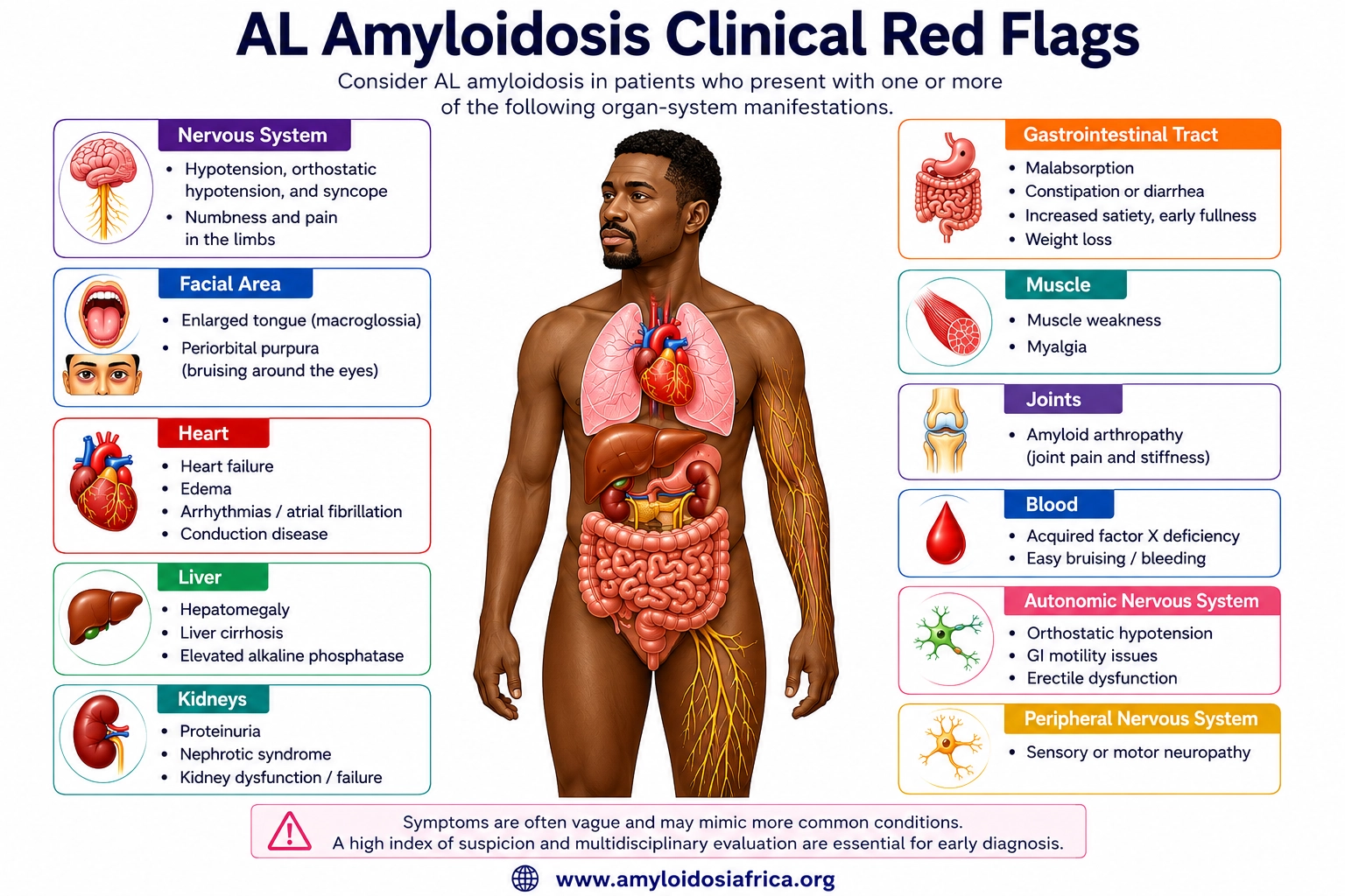
AL amyloidosis is a multisystem disease. Therefore, the pattern and severity of organ involvement can vary substantially between patients.
Commonly Involved Organ Systems
- Heart: restrictive cardiomyopathy, heart failure, arrhythmias and conduction disease
- Kidneys: proteinuria, nephrotic syndrome and progressive kidney dysfunction
- Peripheral nerves: sensory and motor neuropathy
- Autonomic nervous system: orthostatic hypotension, gastrointestinal dysmotility and other autonomic symptoms
- Gastrointestinal tract: altered motility, diarrhea, constipation, early satiety or malabsorption
- Liver: hepatomegaly and abnormal liver-related findings
- Soft tissues: macroglossia, periorbital purpura and other manifestations
How Does AL Amyloidosis Affect the Heart?
When amyloid fibrils accumulate in the myocardium, they make the ventricular walls progressively stiff and may increase the apparent wall thickness on cardiac imaging. Consequently, ventricular relaxation worsens, intracardiac filling pressures rise and cardiac output may eventually fall.
Patients may develop:
- Shortness of breath
- Reduced exercise tolerance
- Peripheral oedema
- Fatigue
- Atrial fibrillation
- Conduction disease
- Hypotension
- Syncope or presyncope
Cardiac AL amyloidosis requires particular urgency. Amyloid infiltration and direct light-chain toxicity can both contribute to cardiac dysfunction. Moreover, cardiac involvement strongly influences prognosis.
What Are the Red Flags for AL Amyloidosis?
No single red flag establishes AL amyloidosis. However, a combination of cardiac, renal, neurological and soft-tissue abnormalities should increase clinical suspicion (1, 2).
Cardiac Clues
- Unexplained increased ventricular wall thickness
- Heart failure with restrictive physiology
- Reduced longitudinal strain with relative apical sparing
- Low or disproportionate ECG voltage in the appropriate clinical context
- Atrial arrhythmias or conduction disease
Systemic Clues
- Proteinuria or nephrotic syndrome
- Macroglossia
- Periorbital purpura
- Peripheral neuropathy
- Autonomic dysfunction
- Unexplained hepatomegaly
Importantly, macroglossia and periorbital purpura are particularly suggestive of AL amyloidosis. Nevertheless, most patients do not need to have either finding for AL amyloidosis to be present.
How Is AL Amyloidosis Diagnosed?
Clinicians need to identify a monoclonal light-chain process and confirm that amyloid is present and correctly typed. Importantly, finding a monoclonal protein alone does not prove AL amyloidosis because monoclonal gammopathy can occur without amyloid disease.
Step 1: Complete Monoclonal Protein Testing
When clinicians suspect AL amyloidosis, they should request:
- Serum free light-chain assay
- Serum immunofixation electrophoresis
- Urine immunofixation electrophoresis
Together, these investigations detect monoclonal light-chain disorders much more effectively than conventional serum protein electrophoresis alone (1).
Important: Clinicians should not use SPEP alone to exclude AL amyloidosis. A complete monoclonal protein screen requires serum free light chains plus serum and urine immunofixation.
Understanding Free Light Chains
The serum free light-chain assay measures circulating free kappa and lambda light chains and calculates the kappa-to-lambda ratio. An abnormal ratio may indicate clonal light-chain production.
However, kidney dysfunction can increase free light-chain concentrations and influence the ratio. Therefore, clinicians should interpret results in the context of kidney function and the laboratory reference range.
Step 2: Confirm Amyloid in Tissue
Clinicians may obtain tissue from abdominal fat, bone marrow or an affected organ, depending on the clinical situation. They can then use Congo red staining to identify amyloid deposits, which classically show apple-green birefringence under polarized light.
Nevertheless, a negative abdominal fat biopsy does not completely exclude AL amyloidosis. If clinical suspicion remains high, clinicians may need to biopsy an affected organ (1).
Step 3: Type the Amyloid
Confirming amyloid in tissue is only one part of the diagnosis. Therefore, clinicians also need to identify the precursor protein because treatment differs fundamentally between AL, ATTR and other amyloid types.
When tissue is available, proteomic analysis using laser microdissection followed by mass spectrometry provides a highly accurate method for determining the amyloid protein (4).
How Is Cardiac Involvement Assessed?
When clinicians diagnose or strongly suspect AL amyloidosis, they should assess the heart because cardiac involvement influences prognosis and treatment decisions. In practice, evaluation may include electrocardiography, echocardiography, cardiac biomarkers and, in selected patients, cardiac magnetic resonance imaging (1, 2).
Electrocardiography
An ECG may show low QRS voltage, pseudoinfarction patterns, atrial fibrillation or conduction abnormalities. However, none of these findings alone has sufficient sensitivity or specificity to diagnose AL cardiac amyloidosis.
Echocardiography
Echocardiography may demonstrate:
- Increased ventricular wall thickness
- Biatrial enlargement
- Diastolic dysfunction
- Reduced longitudinal strain
- Relative apical sparing
- Valve or interatrial septal thickening
- Pericardial effusion
Importantly, these findings can increase suspicion but do not establish the amyloid type.
Cardiac Magnetic Resonance
Cardiac magnetic resonance may show diffuse subendocardial or transmural late gadolinium enhancement, abnormal gadolinium kinetics, elevated native T1 and increased extracellular volume. Although these findings support an infiltrative cardiomyopathy, CMR does not reliably distinguish AL from ATTR by itself (2).
Cardiac Biomarkers
Troponin and natriuretic peptides such as NT-proBNP help clinicians assess cardiac involvement, disease severity and prognosis. Moreover, established AL amyloidosis staging systems incorporate cardiac biomarkers.
Can Bone Scintigraphy Diagnose AL Amyloidosis?
No. Clinicians mainly use PYP, DPD or HMDP bone-avid tracer scintigraphy as part of the non-biopsy diagnostic pathway for ATTR cardiac amyloidosis. Therefore, scintigraphy does not establish a diagnosis of AL amyloidosis.
Importantly, some patients with AL cardiac amyloidosis can show cardiac tracer uptake. Consequently, clinicians must not interpret a positive scan as ATTR-CM until they have completed an appropriate monoclonal protein assessment (5).
Do not diagnose ATTR-CM from scintigraphy alone when a monoclonal protein is present. In that situation, clinicians may need tissue confirmation and definitive amyloid typing.
A Simplified Diagnostic Pathway for Suspected AL Amyloidosis
Clinical suspicion
Cardiac, renal, neurological or systemic red flags
↓
Complete monoclonal protein screen
Serum free light chains + serum immunofixation + urine immunofixation
↓
Monoclonal light-chain abnormality detected
↓
Hematology assessment
↓
Tissue biopsy
Congo red confirmation of amyloid
↓
Definitive amyloid typing
Confirm AL rather than ATTR or another amyloid type
↓
Assess organ involvement and disease stage
Heart, kidneys, nerves and other affected systems
↓
Begin clone-directed treatment
How Is AL Amyloidosis Treated?
Treatment aims to suppress the clonal plasma-cell or B-cell population that produces the amyloidogenic light chain as rapidly and deeply as possible. As a result, treatment can stop further precursor production and allow affected organs to stabilize or recover (3).
Daratumumab-Based Therapy
Daratumumab is a monoclonal antibody that targets CD38 on plasma cells. In the phase 3 ANDROMEDA trial, investigators added subcutaneous daratumumab to bortezomib, cyclophosphamide, and dexamethasone in patients with newly diagnosed systemic AL amyloidosis.
Compared with the regimen without daratumumab, the daratumumab-containing regimen produced substantially higher rates of hematologic complete response and improved major organ deterioration-progression-free survival (3).
Autologous Stem Cell Transplantation
High-dose chemotherapy followed by autologous stem cell transplantation may help carefully selected patients. However, clinicians must assess cardiac involvement, hypotension, kidney dysfunction, frailty and other comorbidities because these factors can substantially increase treatment-related risk.
Therefore, clinicians experienced in AL amyloidosis and plasma-cell disorders should carefully plan treatment and transplant eligibility.
Supportive Cardiac Treatment in AL Amyloidosis
Disease-modifying hematologic treatment does not replace management of heart failure and other cardiac complications. Moreover, patients with AL cardiac amyloidosis may be especially sensitive to hypotension and changes in intravascular volume.
Diuretics
Loop diuretics play a central role in treating congestion. However, clinicians should titrate them carefully to relieve fluid overload while avoiding excessive reduction in preload, symptomatic hypotension and worsening kidney function (1).
Conventional Heart Failure Medications
Beta-blockers, ACE inhibitors, ARBs and ARNIs may cause poor tolerance in some patients because of low blood pressure, restrictive physiology and autonomic dysfunction. Therefore, clinicians should individualize their use rather than automatically applying conventional heart-failure algorithms (1).
Atrial Fibrillation and Thromboembolism
Atrial fibrillation may cause significant symptoms because atrial contraction contributes importantly to ventricular filling in restrictive physiology. In addition, cardiac amyloidosis increases the risk of intracardiac thrombus and systemic thromboembolism.
Because thromboembolic risk is increased, patients with cardiac amyloidosis and atrial fibrillation generally require anticoagulation irrespective of CHA2DS2-VASc score unless contraindicated (1).
How Is Treatment Response Monitored?
Clinicians assess treatment response in two complementary ways: by measuring the hematologic response of the abnormal plasma-cell clone and by evaluating changes in affected organs.
For example, monitoring may include:
- Serum free light chains
- Serum and urine monoclonal protein assessment when appropriate
- NT-proBNP and troponin
- Kidney function and proteinuria
- Symptoms and functional status
- Relevant organ-specific investigations
Importantly, hematologic improvement may occur before an organ response becomes apparent. Therefore, clinicians should interpret laboratory and clinical changes together over time.
AL Amyloidosis in Africa
The true burden of AL amyloidosis across African populations remains insufficiently characterized. Moreover, its symptoms overlap with many common cardiovascular, renal and neurological diseases. Consequently, delayed recognition may be particularly problematic where access to specialized hematology, immunofixation, free light-chain testing, pathology and amyloid typing remains limited.
Therefore, important priorities include:
- Improving awareness of AL-specific red flags
- Expanding access to serum free light-chain testing
- Improving access to serum and urine immunofixation
- Strengthening pathology and Congo red diagnostic capacity
- Developing reliable amyloid-typing pathways
- Establishing rapid referral pathways between cardiology, nephrology and hematology
- Improving access to contemporary clone-directed treatment
- Generating African epidemiological and outcomes data
Key Takeaways
- AL amyloidosis results from amyloidogenic immunoglobulin light chains produced by a clonal plasma-cell or B-cell population.
- The disease can affect the heart, kidneys, peripheral nerves, autonomic nervous system, gastrointestinal tract, liver and soft tissues.
- Cardiac involvement is a major determinant of prognosis and requires timely recognition.
- Proteinuria, nephrotic syndrome, macroglossia and periorbital purpura are important clinical clues.
- A complete monoclonal protein screen includes serum free light chains, serum immunofixation and urine immunofixation.
- Clinicians should not use SPEP alone to exclude AL amyloidosis.
- A monoclonal gammopathy alone does not prove AL amyloidosis.
- When required, clinicians should confirm amyloid in tissue and accurately type the precursor protein.
- A positive PYP, DPD or HMDP scan should not automatically be interpreted as ATTR-CM when a monoclonal protein is present.
- Treatment aims to suppress the abnormal light-chain-producing clone rapidly and deeply.
- Daratumumab-based therapy is an important component of first-line treatment for many patients with newly diagnosed systemic AL amyloidosis.
- Carefully selected patients may be candidates for autologous stem cell transplantation.
- Management of cardiac AL amyloidosis requires close collaboration between hematology and cardiology.
References
- Kittleson MM, Ruberg FL, Ambardekar AV, Brannagan TH 3rd, Cheng RK, Clarke JO, et al. 2023 ACC Expert Consensus Decision Pathway on comprehensive multidisciplinary care for the patient with cardiac amyloidosis. J Am Coll Cardiol. 2023;81(11):1076-1126. doi:10.1016/j.jacc.2022.11.022.
- Garcia-Pavia P, Rapezzi C, Adler Y, Arad M, Basso C, Brucato A, et al. Diagnosis and treatment of cardiac amyloidosis: a position statement of the ESC Working Group on Myocardial and Pericardial Diseases. Eur Heart J. 2021;42(16):1554-1568. doi:10.1093/eurheartj/ehab072.
- Kastritis E, Palladini G, Minnema MC, Wechalekar AD, Jaccard A, Lee HC, et al. Daratumumab-based treatment for immunoglobulin light-chain amyloidosis. N Engl J Med. 2021;385(1):46-58. doi:10.1056/NEJMoa2028631.
- Vrana JA, Gamez JD, Madden BJ, Theis JD, Bergen HR 3rd, Dogan A. Classification of amyloidosis by laser microdissection and mass spectrometry-based proteomic analysis in clinical biopsy specimens. Blood. 2009;114(24):4957-4959. doi:10.1182/blood-2009-07-230722.
- Gillmore JD, Maurer MS, Falk RH, Merlini G, Damy T, Dispenzieri A, et al. Nonbiopsy diagnosis of cardiac transthyretin amyloidosis. Circulation. 2016;133(24):2404-2412. doi:10.1161/CIRCULATIONAHA.116.021612.
Medical Disclaimer
This article provides educational and informational content only and does not replace individualized medical advice, diagnosis or treatment. Qualified healthcare professionals should make diagnostic and treatment decisions according to each patient’s clinical presentation, amyloid type, organ involvement, disease stage, comorbidities, locally applicable guidance and available therapies.

Leave a Reply