



AA Amyloidosis: Causes, Symptoms, Diagnosis and Treatment
AA amyloidosis is a systemic form of amyloidosis that develops when persistent inflammation causes prolonged production of serum amyloid A (SAA). The liver produces SAA as part of the acute inflammatory response. However, when inflammation continues for long periods, SAA may remain persistently elevated. In susceptible individuals, fragments of SAA can misfold, form amyloid fibrils and accumulate in tissues (1, 2).
AA amyloidosis was historically called secondary amyloidosis because another inflammatory, infectious or autoinflammatory disease drives the process. By contrast, AL amyloidosis develops from abnormal immunoglobulin light chains produced by a clonal plasma-cell population, while ATTR amyloidosis results from transthyretin misfolding.
Importantly, the kidneys are the most commonly affected organs in AA amyloidosis. Therefore, clinicians should consider AA amyloidosis when a patient with longstanding inflammatory disease develops unexplained proteinuria, nephrotic syndrome or progressive kidney dysfunction (1).
Key principle: Persistent inflammation drives AA amyloidosis. Therefore, treatment focuses primarily on controlling the underlying inflammatory process and reducing serum amyloid A production.
What Is AA Amyloidosis?
Serum amyloid A is an acute-phase protein that rises rapidly during inflammation. Inflammatory cytokines stimulate the liver to increase SAA production during infection, tissue injury and inflammatory disease.
Normally, SAA levels fall when inflammation resolves. However, persistent inflammatory activity can keep SAA concentrations elevated for months or years. Consequently, prolonged exposure to high SAA concentrations creates conditions that favor AA amyloid fibril formation (1).
Once amyloid fibrils form, they can accumulate extracellularly within organs. Over time, continued deposition may disrupt normal tissue structure and impair organ function.
Chronic inflammation
↓
Inflammatory cytokine activity
↓
Liver increases serum amyloid A production
↓
Persistent SAA elevation
↓
AA amyloid fibril formation
↓
Tissue deposition and organ dysfunction
↓
Inflammatory cytokine activity
↓
Liver increases serum amyloid A production
↓
Persistent SAA elevation
↓
AA amyloid fibril formation
↓
Tissue deposition and organ dysfunction
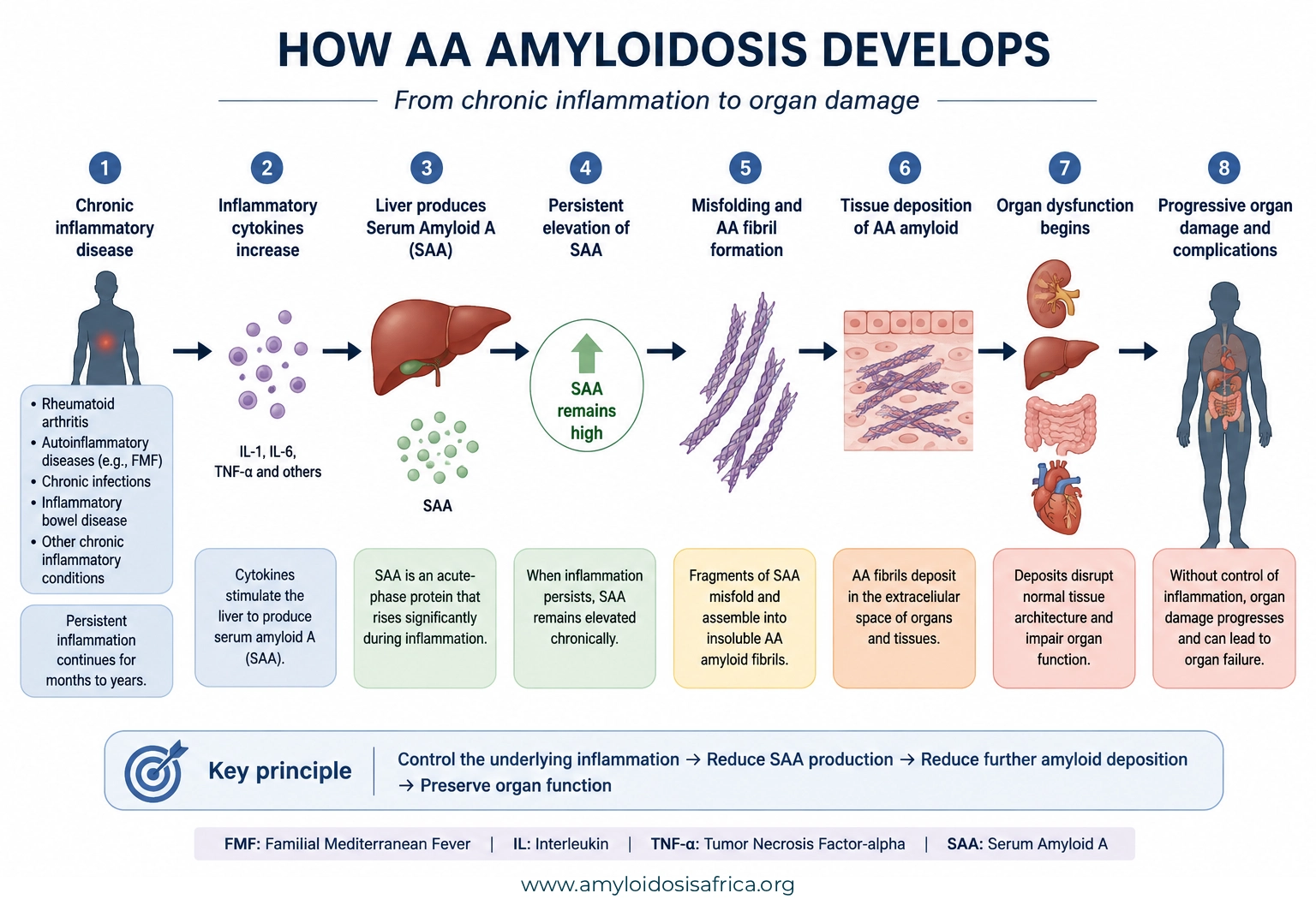
Figure 1. How AA Amyloidosis Develops. Persistent inflammation increases inflammatory cytokines such as IL-1, IL-6, and TNF-α, which stimulate the liver to produce serum amyloid A (SAA). When SAA remains chronically elevated, SAA-derived fragments can form AA amyloid fibrils that deposit in tissues and impair organ function. Controlling the underlying inflammatory disease and suppressing SAA production are therefore central to limiting further amyloid deposition and organ damage.
What Causes AA Amyloidosis?
AA amyloidosis usually develops because another disease produces sustained inflammatory activity. Therefore, identifying the underlying inflammatory driver is a central part of both diagnosis and treatment (2).
Chronic Inflammatory Diseases
Inflammatory arthropathies have historically represented important causes of AA amyloidosis. For example, rheumatoid arthritis and other chronic inflammatory disorders can maintain a persistent acute-phase response when clinicians cannot achieve adequate disease control.
Fortunately, modern anti-inflammatory and biologic treatments have improved control of many inflammatory diseases. As a result, clinicians can often reduce sustained SAA production more effectively than in the past.
Autoinflammatory Diseases
Autoinflammatory syndromes also play an important role. In particular, familial Mediterranean fever (FMF) can cause recurrent or persistent inflammatory activity and, if inadequately controlled, may lead to AA amyloidosis (3).
Therefore, prevention of sustained inflammation forms a major component of FMF management.
Chronic Infections
Persistent infections can also maintain prolonged SAA production. Consequently, clinicians must identify and treat the underlying infectious source whenever infection drives the inflammatory process.
In this setting, simply suppressing inflammatory markers without addressing the infection would not adequately treat the cause of AA amyloidosis.
Other Chronic Inflammatory Conditions
Inflammatory bowel disease and several other chronic inflammatory disorders can also lead to AA amyloidosis. Therefore, clinicians should consider the entire inflammatory history rather than limiting the evaluation to rheumatologic disease alone (1, 2).
Which Organs Can AA Amyloidosis Affect?
AA amyloidosis is a systemic disease, so amyloid deposits can occur in several organs. Nevertheless, its clinical pattern differs from AL and ATTR amyloidosis.
Kidney Involvement in AA Amyloidosis
The kidneys are the dominant clinically affected organs. Renal involvement commonly begins with proteinuria and may progress to nephrotic syndrome, declining glomerular filtration and, eventually, kidney failure (1).
Therefore, clinicians should pay particular attention to renal abnormalities in patients with longstanding inflammatory disease.
Possible warning signs include:
- Proteinuria
- Foamy urine
- Peripheral or generalized edema
- Low serum albumin
- Nephrotic syndrome
- Progressive decline in kidney function
- Kidney failure in advanced disease
Gastrointestinal, Liver and Other Organ Involvement
AA amyloid can also accumulate in the gastrointestinal tract, liver, spleen and other tissues. However, the extent and clinical importance of these deposits vary between patients.
Thus, clinicians should tailor organ assessment to the patient’s symptoms, examination findings and underlying inflammatory disease.
Does AA Amyloidosis Affect the Heart?
Cardiac involvement can occur in AA amyloidosis. However, clinically important amyloid cardiomyopathy is much less characteristic of AA than of AL or ATTR amyloidosis.
Therefore, clinicians should not assume that an amyloid cardiomyopathy represents AA simply because the patient also has chronic inflammation. Instead, they should accurately type the amyloid before making disease-specific treatment decisions.
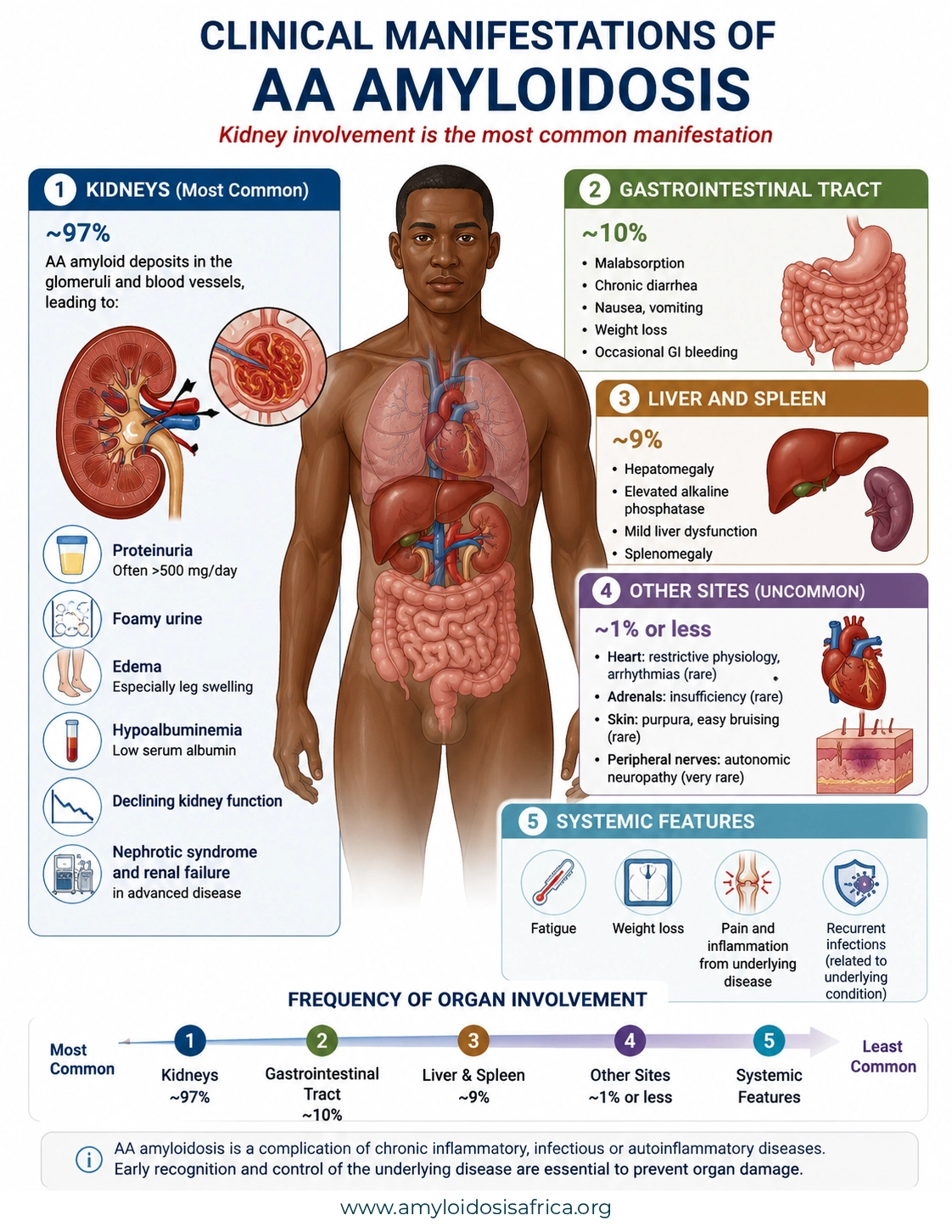
Figure 2. Clinical Manifestations of AA Amyloidosis. AA amyloidosis most commonly affects the kidneys, where amyloid deposition can cause proteinuria, nephrotic syndrome and progressive loss of kidney function. Gastrointestinal and hepatosplenic involvement may also occur, while cardiac, adrenal, skin and peripheral nerve involvement is less common. The pattern and severity of disease vary according to the underlying inflammatory condition, duration of inflammation and timing of treatment. Data source: Australian Amyloidosis Network, adapted from Lachmann HJ et al. Natural History and Outcome in Systemic AA Amyloidosis. New England Journal of Medicine. 2007;356:2361–2371. https://aan.org.au/health-professionals-old/amyloidosis-types/aa/
When Should Clinicians Suspect AA Amyloidosis?
Clinical suspicion should increase when a patient with longstanding inflammatory, infectious or autoinflammatory disease develops otherwise unexplained renal abnormalities.
Chronic inflammatory disease + unexplained proteinuria = consider AA amyloidosis
However, chronic inflammation alone does not establish the diagnosis. Likewise, an elevated SAA concentration shows inflammatory activity but does not prove that amyloid deposits are present.
Therefore, clinicians need a structured diagnostic evaluation when the clinical pattern raises suspicion.
How Is AA Amyloidosis Diagnosed?
Diagnosis requires several complementary steps. First, clinicians assess for organ involvement. Next, they evaluate inflammatory activity. They then confirm amyloid deposition in tissue and, critically, identify the amyloid precursor protein (2).
Step 1: Assess Organ Involvement
When kidney involvement is suspected, clinicians commonly evaluate urinary protein excretion, serum creatinine, estimated glomerular filtration rate and serum albumin.
In addition, symptoms or examination findings may justify gastrointestinal, hepatic or other organ-specific investigations.
Step 2: Assess Inflammatory Activity
C-reactive protein and, where available, serum amyloid A can help clinicians assess the activity of the underlying inflammatory process.
SAA has particular biological relevance because it is the precursor protein for AA amyloid fibrils. However, elevated SAA also occurs in many inflammatory conditions. Therefore, clinicians cannot use an elevated SAA level alone to diagnose AA amyloidosis (1).
Step 3: Confirm Amyloid in Tissue
Tissue biopsy remains central to establishing amyloid deposition. Pathologists typically use Congo red staining to demonstrate amyloid (2).
Clinicians choose the biopsy site according to the patient’s presentation, suspected organ involvement and local diagnostic expertise.
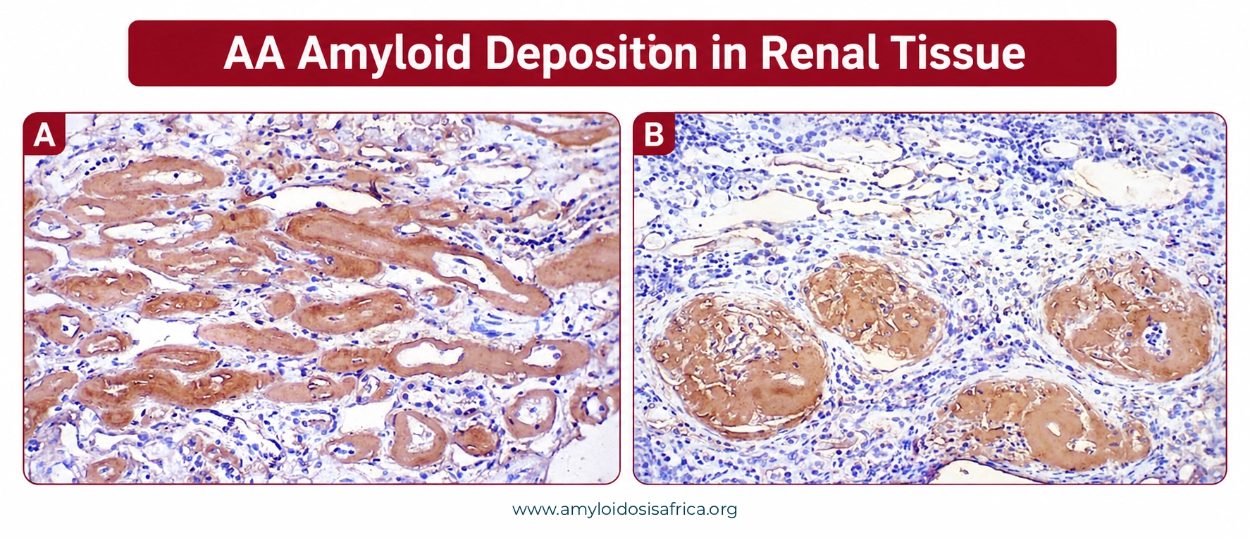
Figure 3. AA Amyloid Deposition in Renal Tissue. Representative kidney histology demonstrating amyloid deposition involving renal structures. Renal involvement is a major manifestation of AA amyloidosis and may lead to proteinuria, nephrotic syndrome, and progressive loss of kidney function.
Step 4: Type the Amyloid Correctly
Finding amyloid in tissue does not complete the diagnostic process. Instead, clinicians must identify the deposited precursor protein because AA, AL, ATTR and other forms of amyloidosis require very different treatments.
Depending on local resources, laboratories may use immunohistochemical techniques or proteomic analysis with mass spectrometry. Importantly, accurate amyloid typing prevents inappropriate treatment and provides the foundation for disease-specific management (4).
Important: Do not assume the amyloid type from the clinical history alone. A patient with chronic inflammation can still have AL, ATTR or another form of amyloidosis.
How Is AA Amyloidosis Treated?
Treatment of AA amyloidosis differs fundamentally from treatment of AL or ATTR amyloidosis.
The primary therapeutic goal is sustained suppression of the inflammatory process that drives SAA production. When treatment controls inflammation effectively, SAA production falls. Consequently, the body has less precursor protein available for continued AA amyloid formation (2).
Therefore, treatment depends heavily on identifying the disease responsible for persistent inflammation.
Treat the Underlying Inflammatory Disease
Clinicians should tailor therapy to the specific inflammatory disorder. For example, inflammatory arthritis, autoinflammatory disease and chronic infection require different therapeutic strategies.
Modern biologic therapies have expanded treatment options for several inflammatory diseases. In selected conditions, cytokine-directed therapies such as IL-6 inhibition can substantially suppress inflammatory activity and SAA production (1).
Nevertheless, clinicians should choose treatment according to the underlying disease rather than treating AA amyloidosis with one universal anti-inflammatory regimen.
Familial Mediterranean Fever and AA Amyloidosis
For familial Mediterranean fever, colchicine remains foundational therapy. Clinicians should begin treatment once they establish the clinical diagnosis of FMF.
However, some patients continue to show inflammatory activity despite appropriate colchicine therapy. In such cases, additional treatment may be necessary. Updated EULAR/PReS recommendations identify strong evidence for IL-1-targeted treatment in appropriate colchicine-resistant patients (3).
When AA amyloidosis complicates FMF, clinicians aim for complete and sustained control of biochemical inflammation. They can monitor this response with SAA or, where SAA testing is unavailable, an alternative inflammatory marker such as CRP (3).
Treat Chronic Infection
When chronic infection drives the inflammatory response, clinicians must identify and effectively treat that infection.
Therefore, treatment focuses on removing or controlling the infectious stimulus rather than simply suppressing inflammatory markers.
Managing Kidney Disease in AA Amyloidosis
Because renal involvement dominates the clinical phenotype, nephrology care often plays a central role in management.
Management may include:
- Monitoring proteinuria and kidney function
- Managing edema and volume status
- Controlling blood pressure
- Managing complications of nephrotic syndrome
- Adjusting therapies according to renal function
- Planning renal replacement therapy if kidney failure develops
Importantly, supportive kidney care does not replace control of the underlying inflammatory disease. Both approaches work together to preserve renal function whenever possible.
Kidney Transplantation
Kidney transplantation can provide an option for carefully selected patients who progress to kidney failure.
Moreover, modern control of the underlying inflammatory disease has improved post-transplant management and reduced the risk associated with ongoing inflammatory activity and recurrent AA deposition (2).
Why Monitoring Serum Amyloid A Matters
SAA is more than an inflammatory biomarker. It is the precursor protein from which AA amyloid fibrils develop.
Therefore, persistent SAA elevation indicates that the biological substrate for further amyloid formation remains present. Conversely, sustained suppression of inflammation reduces SAA production and helps limit continued amyloid deposition.
When available, serial SAA measurements can help clinicians assess inflammatory control over time. However, SAA testing is not universally available. In such settings, CRP may provide a practical alternative marker of inflammatory activity (3).
Treatment target: Achieve sustained control of the underlying inflammatory disease and suppress the acute-phase response as completely as possible.
AA vs AL vs ATTR Amyloidosis
AA, AL and ATTR amyloidosis all involve extracellular amyloid deposition. However, they originate from different precursor proteins and require fundamentally different treatment strategies.
| Feature | AA | AL | ATTR |
|---|---|---|---|
| Precursor protein | Serum amyloid A | Immunoglobulin light chain | Transthyretin |
| Main source | Liver during inflammation | Clonal plasma cells | Liver |
| Main disease driver | Persistent inflammation | Plasma-cell disorder | TTR instability and misfolding |
| Common major organ involvement | Kidneys | Heart and kidneys | Heart ± peripheral/autonomic nerves |
| Main treatment principle | Suppress inflammation and SAA production | Suppress the plasma-cell clone | TTR-directed treatment |
| Hereditary amyloid type? | Usually acquired; however, the underlying autoinflammatory disease may be inherited | No | ATTRv is hereditary; ATTRwt is acquired |
Why typing matters: Amyloidosis is not one disease. Therefore, identifying the deposited precursor protein is essential for choosing the correct treatment.
AA Amyloidosis in Africa
Reliable contemporary epidemiological data that define the burden of AA amyloidosis across African countries remain limited. Therefore, clinicians and researchers should not infer prevalence simply from the number of diagnosed cases.
Nevertheless, the clinical principle remains important. When patients with chronic inflammatory or infectious diseases develop unexplained proteinuria or progressive kidney dysfunction, AA amyloidosis should enter the differential diagnosis.
Priorities for Earlier Recognition in African Settings
Several practical priorities could strengthen recognition and management across African healthcare systems:
- Increase awareness among nephrologists, rheumatologists, infectious-disease physicians and internists
- Recognize unexplained proteinuria in patients with persistent inflammatory disease
- Strengthen renal biopsy and Congo red staining capacity
- Improve access to reliable amyloid typing
- Develop referral pathways linking nephrology, rheumatology, infectious diseases and pathology
- Generate African epidemiological and clinical data on AA amyloidosis
In addition, stronger multidisciplinary pathways could shorten the interval between renal presentation, tissue diagnosis, amyloid typing and treatment of the underlying inflammatory disease.
Key Takeaways About AA Amyloidosis
- AA amyloidosis develops because persistent inflammation causes sustained serum amyloid A production.
- The kidneys are the dominant clinically affected organs.
- Therefore, proteinuria, nephrotic syndrome and progressive kidney dysfunction are important warning signs.
- AA amyloidosis can complicate chronic inflammatory diseases, autoinflammatory syndromes and persistent infections.
- An elevated SAA level indicates inflammatory activity; however, it does not diagnose amyloidosis by itself.
- Clinicians need tissue confirmation and accurate amyloid typing before making disease-specific treatment decisions.
- Treatment primarily aims to suppress the inflammatory process responsible for sustained SAA production.
- For FMF, colchicine remains foundational therapy, while clinicians may consider additional IL-1-targeted therapy when inflammatory activity remains inadequately controlled.
- Patients with advanced renal disease may require renal replacement therapy or, in selected cases, kidney transplantation.
- Finally, AA, AL and ATTR amyloidosis require different treatment strategies. Accurate typing is therefore critical.
Recognize the Link Between Inflammation and AA Amyloidosis
AA amyloidosis is a serious complication of persistent inflammatory activity. However, modern control of the underlying disease can substantially alter its course.
The central clinical challenge is recognizing the connection between chronic inflammation and otherwise unexplained organ dysfunction, particularly renal disease.
Therefore, in a patient with longstanding inflammatory disease, unexplained proteinuria or progressive kidney dysfunction should prompt consideration of AA amyloidosis.
Recognize persistent inflammation. Identify amyloid. Type it correctly. Control the inflammatory driver.
References
- Thorne J, Clark D, Geldenhuys L, et al. Serum Amyloid A Protein–Associated Kidney Disease: Presentation, Diagnosis, and Management. Kidney Med. 2022;4(8):100504. doi:10.1016/j.xkme.2022.100504.
- Mirioglu S, et al. AA Amyloidosis: A Contemporary View. Curr Rheumatol Rep. 2024.
- Ozen S, Sağ E, Oton T, Gül A, Sieiro Santos C, Bayraktar D, et al. EULAR/PReS endorsed recommendations for the management of familial Mediterranean fever (FMF): 2024 update. Ann Rheum Dis. doi:10.1016/j.ard.2025.01.028.
- Leung N, Nasr SH. 2024 Update on Classification, Etiology, and Typing of Renal Amyloidosis: A Review. Am J Kidney Dis. 2024. doi:10.1053/j.ajkd.2024.01.530.
Medical Disclaimer
This article provides educational and informational content only and does not replace individualized medical advice, diagnosis or treatment. Clinicians should individualize evaluation and management according to the underlying inflammatory disorder, organ involvement, comorbidities, locally available investigations and current specialist guidance.

Leave a Reply