



Bone Scintigraphy Imaging of Cardiac Amyloidosis
Bone scintigraphy in cardiac amyloidosis has transformed the diagnosis of transthyretin amyloid cardiomyopathy (ATTR-CM). Technetium-labeled bone-seeking tracers can demonstrate myocardial tracer uptake associated with transthyretin amyloid deposition. Moreover, when clinicians combine imaging findings with appropriate monoclonal protein testing, bone scintigraphy can support a non-invasive diagnosis of ATTR-CM in selected patients (1, 2, 3, 4).
The principal tracers used for cardiac amyloidosis are technetium-99m pyrophosphate (99mTc-PYP), technetium-99m diphosphono-1,2-propanodicarboxylic acid (99mTc-DPD), and technetium-99m hydroxymethylene diphosphonate (99mTc-HMDP). Although nuclear medicine originally developed these agents for skeletal imaging, they can show substantial myocardial uptake in ATTR-CM (2, 3, 5, 7).
However, clinicians must never interpret bone scintigraphy in isolation. In particular, they must evaluate for a monoclonal protein because light-chain (AL) amyloidosis can also involve the heart and requires a fundamentally different diagnostic and treatment pathway (3, 4).
Key principle: Bone scintigraphy can support a non-biopsy diagnosis of ATTR-CM only when clinicians interpret the imaging pattern, SPECT findings, monoclonal protein assessment, and overall clinical context together.
What Is Bone Scintigraphy in Cardiac Amyloidosis?
Bone scintigraphy is a nuclear imaging technique in which a small amount of a technetium-labeled radiotracer is administered intravenously. A gamma camera then detects the distribution of the tracer within the body.
In ATTR-CM, bone-seeking tracers may accumulate within the myocardium. The precise mechanism responsible for preferential tracer binding in transthyretin amyloid deposits remains incompletely understood. Nevertheless, this imaging phenomenon provides powerful diagnostic information when clinicians use standardized acquisition and interpretation protocols.
Importantly, the test does not simply answer whether cardiac amyloid is present. Its greatest clinical value is its ability to support the diagnosis of transthyretin cardiac amyloidosis when clinicians have appropriately excluded AL amyloidosis (3, 4).
Why Is Bone Scintigraphy Important in ATTR-CM?
Historically, clinicians often required endomyocardial biopsy to establish the type of cardiac amyloidosis. However, the development of validated bone scintigraphy pathways has substantially reduced that need.
Gillmore and colleagues demonstrated that moderate or strong cardiac uptake on bone scintigraphy, together with the absence of a monoclonal protein, can provide a highly specific non-biopsy diagnosis of ATTR-CM (3).
As a result, bone scintigraphy now occupies a central position in contemporary diagnostic algorithms for suspected transthyretin cardiac amyloidosis (1, 4, 9).
This is especially important because ATTR-CM may present with nonspecific features such as heart failure with preserved ejection fraction, unexplained increased ventricular wall thickness, atrial fibrillation, conduction disease, or extracardiac red flags. Therefore, scintigraphy usually forms part of a broader pattern-recognition strategy rather than functioning as an isolated test (6).
Bone-Seeking Tracers: PYP, DPD and HMDP
99mTc-PYP
99mTc-pyrophosphate (PYP) is widely used for ATTR-CM evaluation, particularly in the United States. Clinicians generally assess PYP uptake using planar imaging together with SPECT or SPECT/CT. In addition, planar PYP imaging may allow calculation of the heart-to-contralateral chest ratio (2, 5).
99mTc-DPD
99mTc-DPD is commonly used in Europe and other regions. Clinicians compare DPD cardiac uptake with skeletal uptake using a visual grading approach commonly referred to as the Perugini grading system (3, 7).
99mTc-HMDP
99mTc-HMDP is another bone-seeking tracer used in suspected ATTR-CM. Its interpretation follows the same fundamental principles: confirm true myocardial localization, assess uptake intensity, and interpret the result together with monoclonal protein testing and the overall clinical phenotype (4, 5).
Tracer availability differs by country and nuclear medicine service. Therefore, local practice often determines whether PYP, DPD, or HMDP is used.
How Is Bone Scintigraphy Performed?
After intravenous tracer administration, nuclear medicine teams acquire images according to a standardized protocol. The precise timing depends on the tracer and local imaging protocol.
For PYP, imaging may begin approximately one hour after injection. However, if substantial blood-pool activity remains, delayed imaging can help clarify whether apparent cardiac activity represents true myocardial uptake. DPD and HMDP protocols commonly use later acquisition times (5).
The examination typically combines:
- Planar chest imaging
- SPECT imaging of the heart
- SPECT/CT where available
- Visual grading of myocardial uptake
- Quantitative assessment where appropriate
Standardization matters because acquisition timing, image quality, blood-pool activity, region-of-interest placement, and tomographic localization can all influence interpretation (5).
How to Interpret Bone Scintigraphy in Cardiac Amyloidosis
Clinicians should interpret bone scintigraphy systematically. First, they should confirm that apparent cardiac tracer activity truly localizes to the myocardium. Next, they should grade myocardial uptake visually. Finally, quantitative measurements may provide additional support in selected situations.
The nuclear imaging result must then be integrated with the patient’s monoclonal protein assessment and overall clinical probability of cardiac amyloidosis.
Perugini Grade 0
Grade 0 means there is no significant myocardial uptake while normal skeletal uptake remains visible (7).
This pattern makes ATTR-CM less likely. However, if clinical suspicion remains high, clinicians should not automatically stop the diagnostic evaluation. Other investigations, including cardiac magnetic resonance or biopsy in selected circumstances, may still be necessary (1, 4).
Perugini Grade 1
Grade 1 represents myocardial tracer uptake that is less intense than rib uptake (7).
This is an equivocal result. Grade 1 does not satisfy the validated non-biopsy criteria for ATTR-CM. Therefore, patients with persistent suspicion usually require additional evaluation, which may include tissue biopsy and definitive amyloid typing (1, 4, 5).
Perugini Grade 2
Grade 2 represents myocardial uptake approximately equal to rib uptake (7).
When SPECT confirms true myocardial localization and appropriate monoclonal protein testing is negative, Grade 2 uptake can support the non-biopsy diagnosis of ATTR-CM in the appropriate clinical context (3, 4).
Perugini Grade 3
Grade 3 represents intense myocardial tracer uptake that exceeds rib uptake, often with reduced skeletal visualization (7).
Again, this result can support a non-biopsy diagnosis of ATTR-CM when clinicians confirm myocardial localization and find no evidence of a monoclonal protein (3, 4).
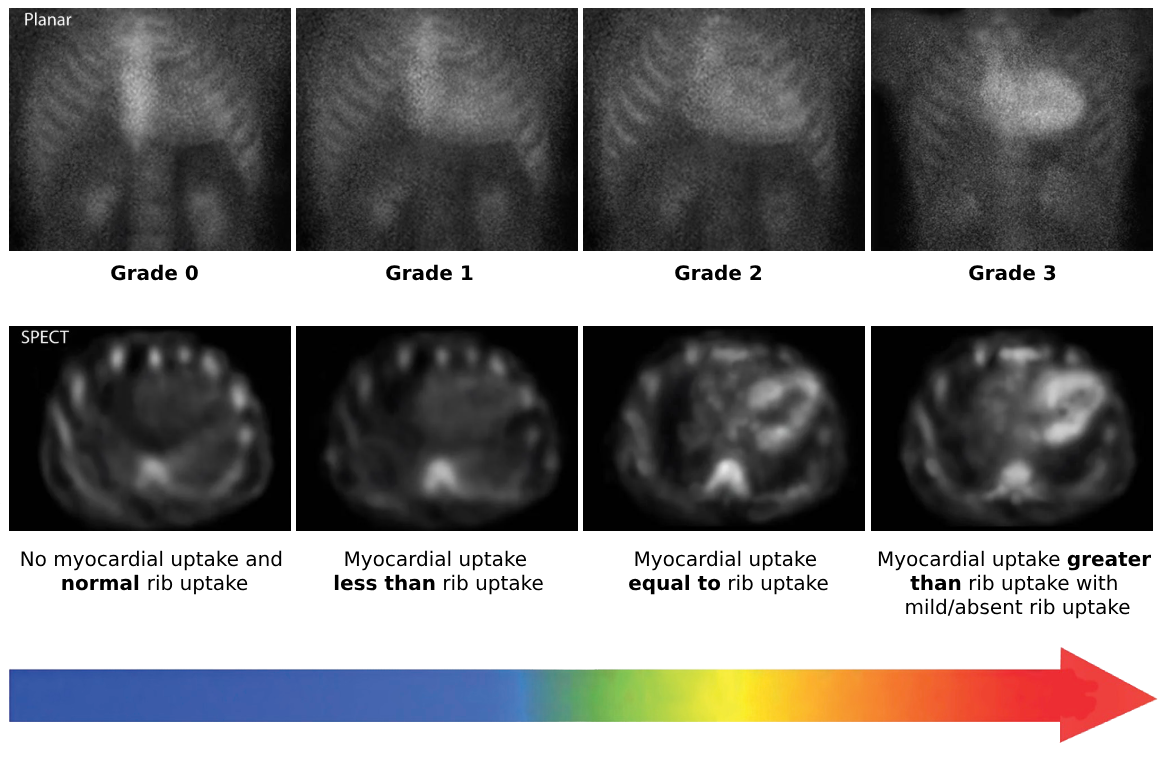
Figure 1. Perugini Grading in Cardiac Amyloidosis. Bone scintigraphy is visually graded from 0 to 3 according to myocardial tracer uptake relative to bone uptake. Grade 0 shows no myocardial uptake; Grade 1 shows uptake less than rib uptake; Grade 2 shows myocardial uptake similar to rib uptake; and Grade 3 shows myocardial uptake greater than rib uptake with reduced or absent skeletal uptake. SPECT should be used to confirm that apparent cardiac activity represents true myocardial uptake rather than blood-pool or overlapping bone activity.
Why Is SPECT Essential in Bone Scintigraphy?
Planar imaging alone cannot always determine whether radiotracer activity lies within the myocardium. For example, persistent blood-pool activity can project over the cardiac silhouette and mimic myocardial uptake.
SPECT is therefore essential for confirming myocardial localization. It provides tomographic images that help separate true myocardial tracer retention from blood-pool activity and nearby skeletal structures (2, 5).
SPECT may also help identify potential confounders such as:
- Blood-pool activity
- Sternal uptake
- Rib uptake overlapping the cardiac silhouette
- Rib fractures
- Other focal bone abnormalities
Where available, SPECT/CT further improves anatomical localization by combining functional tracer imaging with CT anatomy.
Clinical takeaway: A bright heart on a planar image is not enough. The interpreter should confirm true myocardial uptake tomographically before using the scan to support an ATTR-CM diagnosis.
Quantitative Assessment and the H/CL Ratio
With 99mTc-PYP, clinicians may supplement visual assessment with the heart-to-contralateral chest ratio (H/CL ratio) (2, 5).
To calculate the ratio, the interpreter draws a region of interest over the heart on an anterior planar image. An identical region is then mirrored over the contralateral chest.
H/CL ratio = Mean counts in heart ROI ÷ Mean counts in contralateral chest ROI
Historically, an H/CL ratio of ≥1.5 at approximately one hour has supported a positive PYP study when the overall imaging and clinical context are appropriate (2, 5).
However, clinicians should not use the H/CL ratio as a stand-alone diagnostic criterion. Blood-pool activity can increase planar counts without representing genuine myocardial uptake. Therefore, SPECT confirmation remains critical.
Moreover, a numerical threshold should never override discordant tomographic findings or an abnormal monoclonal protein assessment.
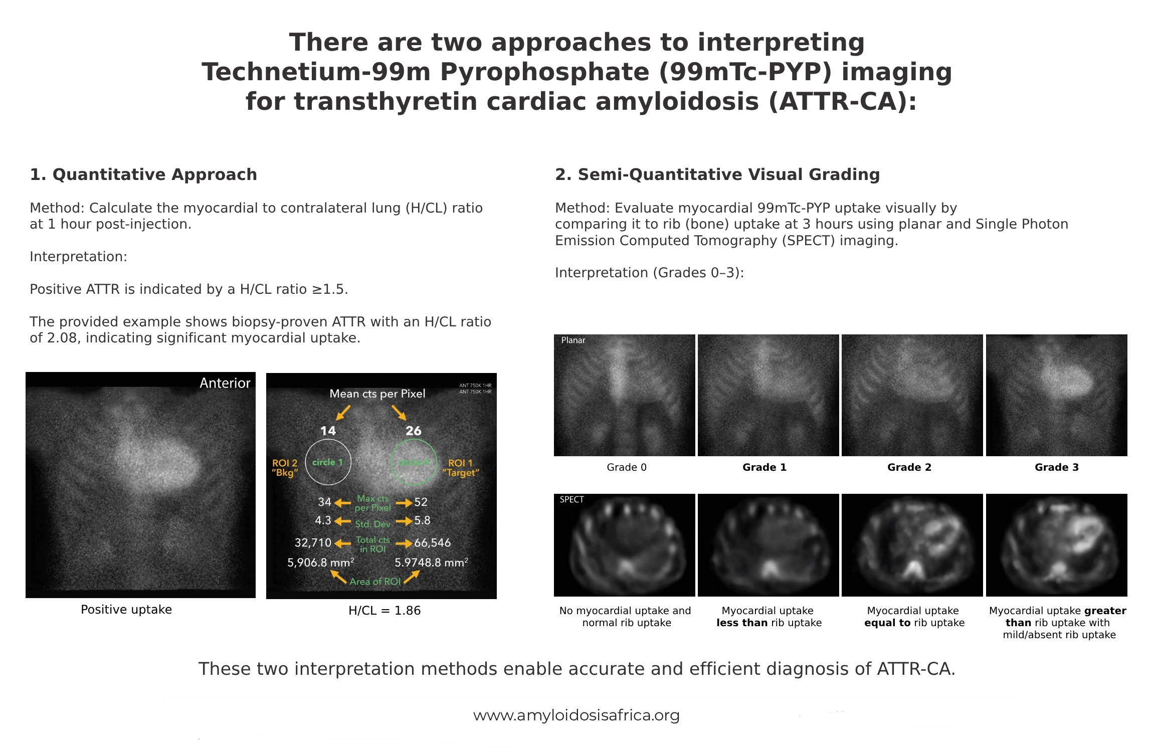
Figure 3. Interpretation of 99mTc-PYP Imaging in Transthyretin Cardiac Amyloidosis. PYP scintigraphy can be assessed using quantitative and semi-quantitative approaches. The quantitative method uses the heart-to-contralateral chest (H/CL) ratio on planar imaging, while visual grading compares myocardial tracer uptake with rib uptake from Grade 0 to Grade 3. SPECT should confirm true myocardial localization, and imaging findings must be interpreted together with monoclonal protein testing before ATTR-CM is diagnosed.
Why Must AL Amyloidosis Be Excluded?
This is one of the most important principles in the entire diagnostic pathway.
Bone-seeking tracer uptake is strongly associated with ATTR-CM. However, cardiac uptake can also occur in AL amyloidosis. Therefore, clinicians cannot safely diagnose ATTR-CM from scintigraphy alone (3, 4).
Every patient undergoing evaluation for suspected cardiac amyloidosis should have an appropriate monoclonal protein assessment that includes:
- Serum free light-chain assay
- Serum immunofixation electrophoresis
- Urine immunofixation electrophoresis
Together, these tests assess for evidence of a plasma-cell disorder that could indicate AL amyloidosis (3, 4).
Important: Serum protein electrophoresis alone is not an adequate monoclonal protein screen for this diagnostic pathway. Serum and urine immunofixation together with serum free light chains are required.
If any monoclonal protein test is abnormal, clinicians should not label the patient as ATTR-CM from bone scintigraphy alone. Instead, further assessment is necessary to exclude AL amyloidosis and determine the actual amyloid type (3, 4).
The Non-Biopsy Diagnostic Pathway for ATTR-CM
The validated non-biopsy diagnostic pathway represents one of the most important advances in cardiac amyloidosis diagnosis (3).
In the appropriate clinical setting, ATTR-CM can be diagnosed without endomyocardial biopsy when the following elements are present:
- There is clinical or imaging suspicion of cardiac amyloidosis.
- Bone scintigraphy demonstrates Grade 2 or Grade 3 myocardial uptake.
- SPECT or SPECT/CT confirms true myocardial tracer localization.
- Serum free light chains, serum immunofixation, and urine immunofixation do not demonstrate evidence suggesting a monoclonal plasma-cell disorder.
When these criteria are satisfied, the findings support the non-invasive diagnosis of ATTR-CM (3, 4).
After ATTR-CM is established, clinicians should perform TTR genetic testing to distinguish hereditary ATTR (ATTRv) from wild-type ATTR (ATTRwt) (1, 4).
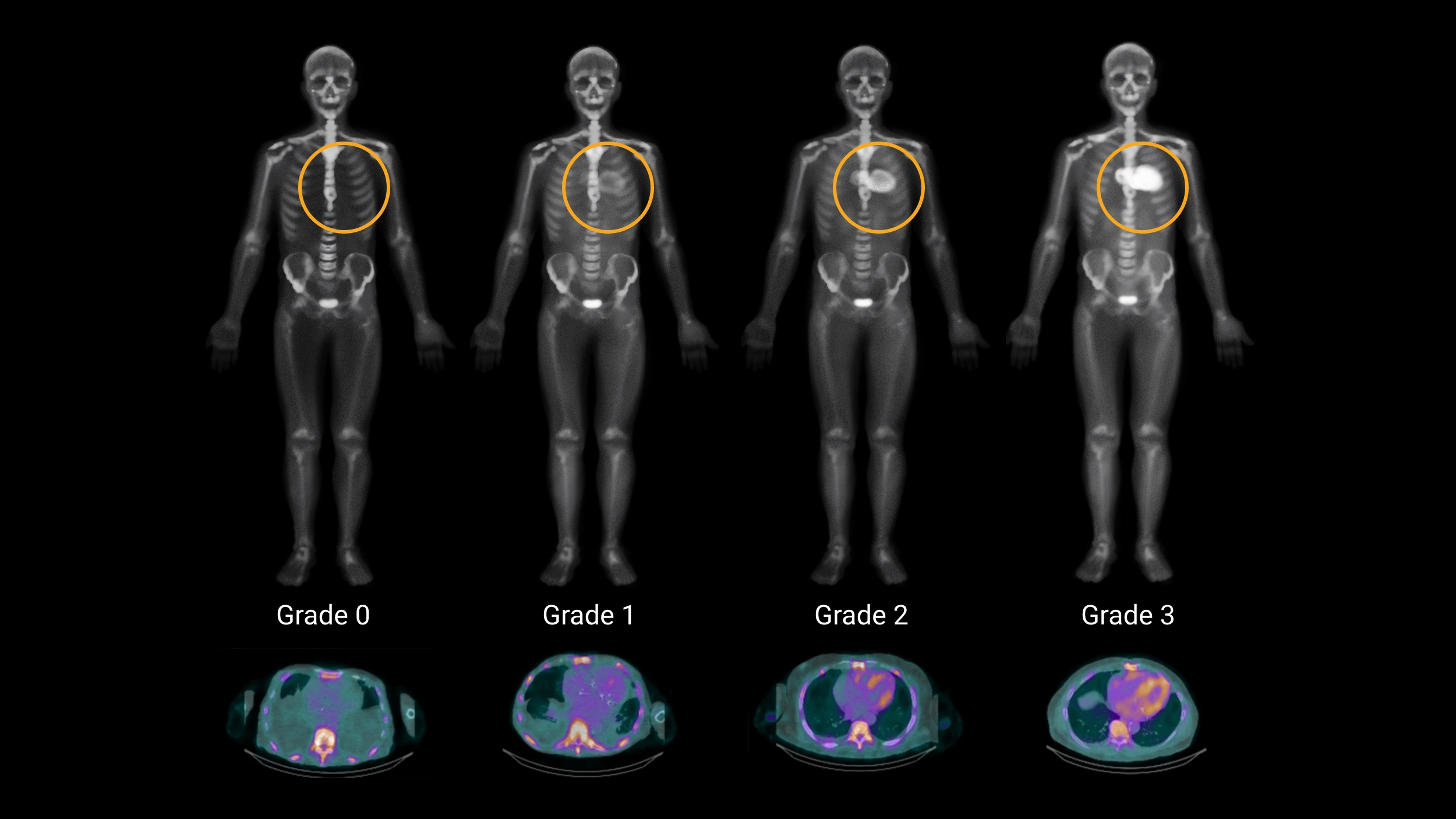
Figure 2. Increasing Myocardial Tracer Uptake on Bone Scintigraphy. Representative imaging illustrates the progression from Grade 0, with no significant myocardial uptake, through Grades 1 and 2 to intense Grade 3 cardiac uptake. The visual grade forms part of the diagnostic assessment for suspected ATTR-CM and must be interpreted together with tomographic confirmation and monoclonal protein testing.
When Is Biopsy Still Required?
Bone scintigraphy has reduced the need for endomyocardial biopsy. Nevertheless, biopsy remains essential in several important situations.
Clinicians should consider tissue confirmation when:
- Monoclonal protein testing is abnormal
- Scintigraphy shows Grade 1 uptake
- Scintigraphy and other clinical findings are discordant
- The scan is negative despite high clinical suspicion
- The amyloid type remains uncertain
- Another amyloid precursor protein needs to be excluded
When biopsy demonstrates amyloid, accurate amyloid typing remains essential. Congo red positivity confirms amyloid deposition, but it does not determine whether the deposits are ATTR, AL, AA, or another amyloid type (4).
Common Pitfalls in Bone Scintigraphy Interpretation
Bone scintigraphy is powerful, but several technical and biological factors can produce misleading results. Therefore, interpretation should always follow a standardized workflow.
Blood-Pool Activity
Residual radiotracer within the cardiac chambers can mimic myocardial uptake, particularly on early planar images. SPECT helps differentiate blood-pool signal from actual myocardial retention (5).
Rib and Sternal Uptake
Bone uptake can overlap the cardiac silhouette. Therefore, rib fractures, focal skeletal abnormalities, and sternal activity may create a false impression of myocardial tracer retention (5).
AL Amyloidosis
AL cardiac amyloidosis can show bone-seeking tracer uptake. Consequently, moderate or strong uptake does not independently prove ATTR-CM (3).
This is precisely why monoclonal protein testing forms an integral part of scan interpretation rather than a separate optional investigation.
Equivocal Grade 1 Uptake
Grade 1 uptake cannot establish ATTR-CM non-invasively. Therefore, if clinical suspicion persists, clinicians need additional investigations (4, 5).
Negative Bone Scintigraphy Despite Clinical Suspicion
A Grade 0 scan makes ATTR-CM less likely. However, no diagnostic test should be interpreted without considering pre-test probability.
When echocardiography, cardiac magnetic resonance, clinical red flags, or family history remain strongly suggestive of amyloidosis, clinicians may need to continue the diagnostic evaluation despite a negative bone scan (1, 4).
Bone Scintigraphy Compared With Echocardiography and CMR
Echocardiography, cardiac magnetic resonance (CMR), and bone scintigraphy answer different but complementary diagnostic questions.
Echocardiography
Echocardiography often raises the initial suspicion of cardiac amyloidosis. Findings may include increased ventricular wall thickness, restrictive physiology, atrial enlargement, reduced tissue Doppler velocities, abnormal global longitudinal strain, and relative apical sparing.
Cardiac Magnetic Resonance
CMR provides detailed myocardial tissue characterization. Late gadolinium enhancement, native T1 mapping, and extracellular volume assessment can demonstrate an infiltrative myocardial process and quantify expansion of the extracellular space.
Bone Scintigraphy
Bone scintigraphy adds something fundamentally different. When myocardial tracer uptake is sufficiently intense and AL amyloidosis has been appropriately excluded, scintigraphy can identify a pattern that is highly characteristic of ATTR-CM and may eliminate the need for myocardial biopsy (3, 4).
Therefore, clinicians should view these imaging modalities as complementary rather than competing tests.
Bone Scintigraphy in Cardiac Amyloidosis in Africa
Bone scintigraphy could play an important role in improving ATTR-CM diagnosis across Africa. However, access to nuclear medicine services, appropriate radiotracers, SPECT or SPECT/CT systems, and experienced interpretation varies between healthcare settings.
In addition, an effective non-biopsy ATTR-CM pathway depends on more than access to the scan itself. Laboratories must also be able to perform the monoclonal protein investigations required to evaluate for AL amyloidosis.
Priorities for African Diagnostic Pathways
Important areas for development include:
- Improving awareness of appropriate indications for bone scintigraphy
- Expanding access to PYP, DPD, or HMDP where feasible
- Strengthening SPECT and SPECT/CT capacity
- Standardizing image acquisition and interpretation
- Ensuring access to serum free light-chain testing
- Ensuring access to serum and urine immunofixation
- Developing referral pathways for equivocal or discordant cases
- Improving access to tissue biopsy and accurate amyloid typing when required
- Training nuclear medicine physicians, cardiologists, and multidisciplinary amyloidosis teams
A standardized approach is particularly important in resource-constrained settings because an incorrect ATTR diagnosis could delay recognition and treatment of AL amyloidosis.
A Practical Bone Scintigraphy Checklist
When interpreting bone scintigraphy in a patient with suspected cardiac amyloidosis, clinicians should ask:
- Was an appropriate bone-seeking tracer used?
- Was the scan acquired using a standardized protocol?
- Is apparent cardiac activity truly myocardial on SPECT or SPECT/CT?
- What is the Perugini visual grade?
- If PYP was used, does quantitative assessment support the visual interpretation?
- Could blood-pool or skeletal activity explain the finding?
- Were serum free light chains performed?
- Were serum and urine immunofixation performed?
- Is there any evidence of a monoclonal protein?
- Do the nuclear imaging findings fit the echocardiographic, CMR, and clinical phenotype?
- If ATTR-CM is established, has TTR genetic testing been arranged?
Key Takeaways
- Bone scintigraphy in cardiac amyloidosis is central to the non-invasive diagnosis of ATTR-CM.
- The principal tracers are 99mTc-PYP, 99mTc-DPD, and 99mTc-HMDP.
- Planar imaging alone is insufficient when myocardial localization is uncertain.
- SPECT or SPECT/CT should confirm true myocardial uptake.
- Perugini Grade 0 represents no myocardial uptake.
- Grade 1 uptake is equivocal and does not establish ATTR-CM.
- Grade 2 and Grade 3 uptake can support a non-biopsy ATTR-CM diagnosis when the full diagnostic criteria are satisfied.
- Monoclonal protein testing is essential in the diagnostic pathway.
- A positive bone scan does not independently exclude AL amyloidosis.
- An abnormal monoclonal protein assessment means ATTR-CM should not be diagnosed from scintigraphy alone.
- The H/CL ratio can support PYP interpretation but should not replace visual assessment and SPECT confirmation.
- Biopsy remains important for equivocal, discordant, or monoclonal-protein-positive cases.
- After ATTR-CM is diagnosed, TTR genetic testing distinguishes ATTRv from ATTRwt.
Conclusion
Bone scintigraphy has fundamentally changed the diagnostic pathway for transthyretin cardiac amyloidosis. PYP, DPD, and HMDP imaging can identify characteristic myocardial tracer uptake and, when clinicians combine these findings with SPECT confirmation and negative monoclonal protein testing, can establish ATTR-CM without endomyocardial biopsy in many appropriately selected patients (3, 4).
However, the strength of the test depends on correct interpretation. A bone scan should never be read in isolation. Planar findings, SPECT localization, visual grade, quantitative measurements, monoclonal protein testing, and the overall clinical phenotype must all be considered together.
The essential principle is straightforward: confirm myocardial uptake, evaluate for AL amyloidosis, and then determine whether the patient meets the validated non-biopsy pathway for ATTR-CM.
References
- Kittleson MM, Maurer MS, Ambardekar AV, et al. Cardiac amyloidosis: evolving diagnosis and management: a scientific statement from the American Heart Association. Circulation. 2020;142(1).
- Bokhari S, Castaño A, Pozniakoff T, Deslisle S, Latif F, Maurer MS. 99mTc-pyrophosphate scintigraphy for differentiating light-chain cardiac amyloidosis from transthyretin-related familial and senile cardiac amyloidoses. Circulation: Cardiovascular Imaging. 2013;6(2):195–201. https://doi.org/10.1161/CIRCIMAGING.112.000132
- Gillmore JD, Maurer MS, Falk RH, et al. Nonbiopsy diagnosis of cardiac transthyretin amyloidosis. Circulation. 2016;133(24):2404–2412. https://doi.org/10.1161/CIRCULATIONAHA.116.021612
- Garcia-Pavia P, Rapezzi C, Adler Y, et al. Diagnosis and treatment of cardiac amyloidosis: a position statement of the ESC Working Group on Myocardial and Pericardial Diseases. European Heart Journal. 2021;42(16):1554–1568.
- Dorbala S, Bokhari S, Miller E, Bullock-Palmer R, Soman P, Thompson R. ASNC Practice Points: 99mTechnetium-pyrophosphate imaging for transthyretin cardiac amyloidosis. American Society of Nuclear Cardiology. 2019.
- Ruberg FL, Grogan M, Hanna M, Kelly JW, Maurer MS. Transthyretin amyloid cardiomyopathy: JACC state-of-the-art review. Journal of the American College of Cardiology. 2019;73(22):2872–2891. https://doi.org/10.1016/j.jacc.2019.04.003
- Perugini E, Guidalotti PL, Salvi F, et al. Noninvasive etiologic diagnosis of cardiac amyloidosis using 99mTc-3,3-diphosphono-1,2-propanodicarboxylic acid scintigraphy. Journal of the American College of Cardiology. 2005;46(6):1076–1084. https://doi.org/10.1016/j.jacc.2005.05.073
- Castaño A, Haq M, Narotsky DL, et al. Multicenter study of planar technetium 99m pyrophosphate cardiac imaging: predicting survival for patients with ATTR cardiac amyloidosis. JAMA Cardiology. 2016;1(8):880–889. https://doi.org/10.1001/jamacardio.2016.2839
- Maurer MS, Bokhari S, Damy T, et al. Expert consensus recommendations for the suspicion and diagnosis of transthyretin cardiac amyloidosis. Circulation: Heart Failure. 2019;12(9):e006075. https://doi.org/10.1161/CIRCHEARTFAILURE.119.006075
Medical Disclaimer
This article provides educational and informational content only and does not replace individualized medical advice, diagnosis, specialist nuclear medicine interpretation, or locally applicable imaging protocols. Clinicians should interpret bone scintigraphy together with the patient’s clinical context, laboratory assessment, echocardiography, cardiac magnetic resonance, and additional investigations when indicated.

[…] Bone Scintigraphy Imaging […]