



Biopsy in Cardiac Amyloidosis: Tissue Diagnosis, Congo Red Staining and Amyloid Typing
Biopsy in cardiac amyloidosis remains an important diagnostic tool even though many patients with transthyretin cardiac amyloidosis (ATTR-CM) can now be diagnosed without cardiac tissue sampling. Biopsy provides direct evidence of amyloid deposition and, crucially, allows the amyloid precursor protein to be identified (1, 2).
The histologic hallmark of amyloidosis is extracellular deposition of amyloid fibrils that bind Congo red and demonstrate characteristic birefringence under polarized light. However, identifying amyloid deposits is only the first step. Clinicians must also determine whether the deposits are composed of immunoglobulin light chains, transthyretin, serum amyloid A, or another amyloidogenic protein (1, 2).
This distinction is critical because the major forms of cardiac amyloidosis require completely different treatments. Therefore, a biopsy report stating only that “amyloid is present” is incomplete when the precursor protein has not been identified.
Key principle: Biopsy confirms amyloid deposition, but accurate amyloid typing determines the disease subtype and guides treatment.
Why Is Biopsy Used in Cardiac Amyloidosis?
Modern imaging has substantially reduced the need for endomyocardial biopsy in ATTR-CM. In appropriately selected patients, Grade 2 or Grade 3 myocardial uptake on bone scintigraphy together with a negative monoclonal protein screen can establish ATTR-CM without tissue biopsy (1, 2).
However, biopsy remains essential when the non-invasive diagnostic pathway cannot confidently establish the amyloid type.
Biopsy may be required when:
- A monoclonal protein is detected in a patient with suspected cardiac amyloidosis
- Bone scintigraphy is negative or equivocal despite strong clinical suspicion
- Bone scintigraphy is unavailable
- Imaging, laboratory findings, and clinical features are discordant
- AL amyloidosis is suspected
- The amyloid precursor protein cannot otherwise be determined
- Coexisting ATTR and AL amyloidosis is suspected
The ACC specifically identifies endomyocardial biopsy as appropriate when clinical suspicion remains high despite negative or equivocal scintigraphy, when a monoclonal protein is present and cardiac amyloidosis remains suspected, or when cardiac scintigraphy is unavailable (1).
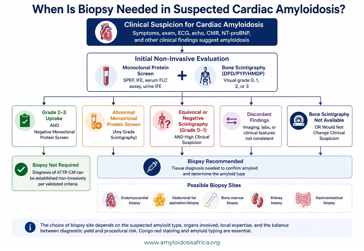
Figure 1. When Is Biopsy Needed in Suspected Cardiac Amyloidosis? Initial evaluation combines monoclonal protein testing with bone scintigraphy. In appropriately selected patients with grade 2–3 cardiac uptake and no evidence of a monoclonal protein, ATTR-CM may be diagnosed non-invasively. Biopsy is generally required when a monoclonal protein is detected or when imaging and clinical findings are equivocal or discordant. When tissue is obtained, Congo red staining confirms amyloid deposition, followed by accurate amyloid typing to establish the amyloid type.
Where Can Amyloid Be Biopsied?
Amyloid can be demonstrated in the affected organ or in a surrogate tissue site. The optimal biopsy site depends on the suspected amyloid type, organs involved, local expertise, and the balance between diagnostic yield and procedural risk.
Common biopsy sites include:
- Endomyocardial biopsy
- Abdominal fat pad aspiration or biopsy
- Bone marrow biopsy
- Kidney biopsy
- Gastrointestinal biopsy
- Salivary gland biopsy
- Other clinically involved organs
Biopsy of an involved organ generally provides a higher diagnostic yield than biopsy of a surrogate site. However, it may also carry greater procedural risk.
Endomyocardial Biopsy
Endomyocardial biopsy directly samples myocardial tissue and remains the definitive invasive method for confirming cardiac amyloid deposition.
According to the ESC, cardiac amyloidosis is definitively confirmed when amyloid deposits are identified in endomyocardial tissue by Congo red staining, irrespective of the degree of ventricular wall thickness (2).
The procedure is usually performed through venous access, commonly from the right side of the heart, with small myocardial tissue samples obtained from the interventricular septum.
Endomyocardial biopsy is particularly valuable when the heart is the dominant affected organ and extracardiac biopsy does not establish the diagnosis.
When Is Endomyocardial Biopsy Particularly Useful?
- Strong suspicion of cardiac amyloidosis with an abnormal monoclonal protein screen
- Negative or equivocal bone scintigraphy despite convincing cardiac imaging
- Suspected simultaneous ATTR and AL cardiac amyloidosis
- Failure of less-invasive biopsy sites to demonstrate amyloid
- Need for definitive characterization of cardiac pathology
Important: A positive extracardiac biopsy can establish systemic amyloidosis, but it does not automatically prove that unexplained cardiac disease is caused by the same amyloid type when clinical findings remain discordant.
Abdominal Fat Pad Aspiration or Biopsy
Abdominal fat sampling is attractive because it is minimally invasive and can often be performed in an outpatient setting. Amyloid may be demonstrated within small vessels and connective tissue in subcutaneous fat.
However, diagnostic sensitivity differs substantially according to amyloid type. The ACC reports approximate sensitivities of 84% for AL cardiac amyloidosis, 45% for ATTRv-CM, and only 15% for ATTRwt-CM (1).
Therefore, a positive fat pad biopsy is useful, but a negative result does not exclude cardiac amyloidosis.
Diagnostic pitfall: A negative abdominal fat pad biopsy should never be used by itself to rule out AL-CM or ATTR-CM when clinical suspicion remains high.
Bone Marrow Biopsy in Suspected AL Amyloidosis
Bone marrow biopsy plays a different but complementary role in suspected AL amyloidosis. It assesses the underlying plasma-cell or B-cell clone responsible for producing the amyloidogenic immunoglobulin light chain.
Amyloid deposits may also be identified directly in the bone marrow. However, the ACC reports that the sensitivity of detecting amyloid deposits in bone marrow in systemic AL amyloidosis is approximately 69% (1).
Consequently, a bone marrow biopsy may establish the clonal plasma-cell disorder even when amyloid itself is not identified in that sample.
When AL amyloidosis is suspected, bone marrow evaluation can also help distinguish AL associated with a small plasma-cell clone from multiple myeloma or less-common B-cell lymphoproliferative disorders (1).
Biopsy of an Affected Organ
If a less-invasive surrogate biopsy is negative but clinical suspicion remains high, biopsy of an affected organ may provide the highest diagnostic yield.
Kidney
Renal biopsy may be appropriate in patients with significant proteinuria, nephrotic syndrome, or unexplained renal dysfunction when renal amyloidosis is suspected. Kidney biopsy can demonstrate both the presence and distribution of amyloid within glomeruli, vessels, and interstitial structures.
Gastrointestinal Tract
Endoscopic biopsy can demonstrate gastrointestinal amyloid deposition, particularly when patients have compatible gastrointestinal symptoms. The ACC notes that Congo red staining should be specifically requested because amyloid may otherwise be missed on routine histology (1).
Other Sites
Minor salivary gland, liver, nerve, skin, or other clinically affected tissues may also be sampled in selected patients. The choice should be guided by the suspected amyloid type, clinical manifestations, procedural safety, and local expertise.
Congo Red Staining: How Amyloid Is Identified
The classic histologic method for identifying amyloid is Congo red staining.
Amyloid fibrils bind Congo red because of their characteristic beta-sheet structure. Under ordinary light microscopy, amyloid deposits appear red or salmon-colored. When viewed under polarized light, Congo red-positive deposits demonstrate characteristic birefringence (2).
This property remains a cornerstone of pathologic diagnosis.

Figure 2. Endomyocardial Biopsy in Cardiac Amyloidosis. Endomyocardial biopsy involves advancing a biopsy catheter through the venous system into the right ventricle to obtain small myocardial tissue samples. Histological examination with Congo red staining can confirm amyloid deposition, while subsequent amyloid typing, preferably using mass spectrometry where available, identifies the amyloid protein and guides appropriate treatment.
Why Must Amyloid Be Typed?
Congo red staining answers one question:
Is amyloid present in the tissue?
It does not answer the equally important second question:
Which protein formed the amyloid?
Accurate typing distinguishes AL, ATTR, AA, and less-common amyloid types. This distinction directly determines treatment.
For example, a patient with ATTR-CM and coincidental monoclonal gammopathy of undetermined significance (MGUS) could easily be misclassified as having AL amyloidosis if clinicians assume that the monoclonal protein explains the tissue deposits.
Therefore, amyloid typing is essential whenever the precursor protein is uncertain (1).
Mass Spectrometry for Amyloid Typing
Mass spectrometry-based proteomic analysis is considered the gold standard for tissue amyloid typing (1).
The technique commonly uses liquid chromatography with tandem mass spectrometry (LC-MS/MS). Amyloid-containing tissue is analyzed to identify the proteins contained within the deposits.
The ACC reports a sensitivity of approximately 88% and specificity of approximately 96% for mass spectrometry-based tissue diagnosis (1).
Although LC-MS/MS is not available in every hospital, Congo red-positive tissue blocks can often be transferred to an experienced reference laboratory for analysis.
Immunohistochemistry and Other Typing Methods
Experienced pathology laboratories may also use immunohistochemistry or immunogold immunoelectron microscopy to identify amyloid precursor proteins.
However, interpretation can be technically challenging. Therefore, mass spectrometry is preferred when available, particularly when the clinical picture and immunostaining results are discordant.
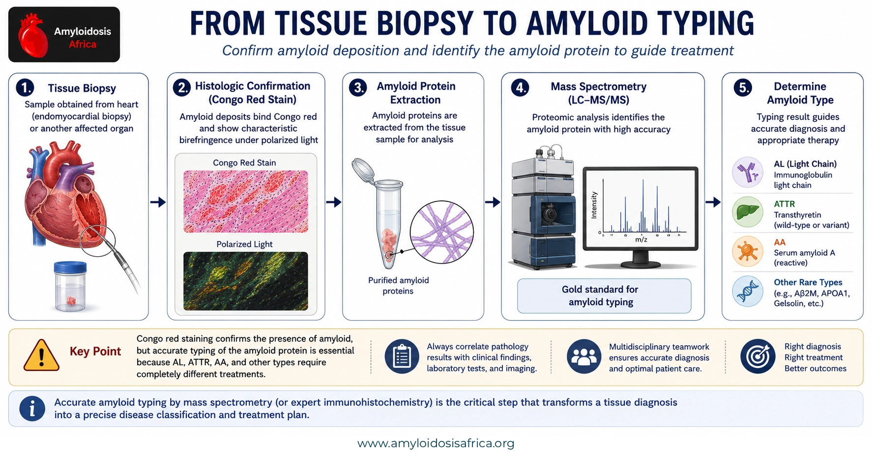
Figure 3. From Tissue Biopsy to Amyloid Typing. Tissue biopsy can confirm amyloid deposition using Congo red staining, with characteristic birefringence under polarized light. Once amyloid is demonstrated, accurate typing is essential to identify the precursor protein and distinguish AL, ATTR, AA, and other amyloid types. Proteomic analysis using liquid chromatography–tandem mass spectrometry (LC-MS/MS) is a highly reliable method for amyloid typing and helps establish the correct diagnosis and guide disease-specific treatment.
Biopsy in Suspected AL Amyloidosis
Unlike the validated non-biopsy pathway available for ATTR-CM, diagnosis of AL amyloidosis generally requires tissue evidence of amyloid together with demonstration of the underlying clonal plasma-cell or B-cell disorder (1).
A common practical strategy is to begin with less-invasive sampling, such as abdominal fat aspiration and bone marrow biopsy. If these specimens are negative but clinical suspicion remains strong, biopsy of the clinically involved organ should be pursued.
When cardiac involvement is dominant and surrogate biopsy is unrevealing, endomyocardial biopsy may be necessary.
When Is Biopsy Needed in ATTR Cardiac Amyloidosis?
Many patients with ATTR-CM do not need a biopsy because contemporary bone scintigraphy can establish the diagnosis non-invasively when appropriate criteria are satisfied.
However, biopsy should still be considered when:
- The monoclonal protein screen is abnormal
- Scintigraphy shows Grade 1 uptake
- Scintigraphy is negative despite strong clinical suspicion
- The imaging phenotype is atypical
- There is concern for another amyloid type
- The result would materially change treatment
Practical distinction: A patient with Grade 2–3 bone scintigraphy and a negative monoclonal protein screen may meet criteria for non-biopsy ATTR-CM diagnosis. A patient with the same scan but an abnormal monoclonal protein screen does not.
Does a Negative Biopsy Exclude Amyloidosis?
Not necessarily.
The diagnostic significance of a negative biopsy depends heavily on the tissue sampled. Surrogate sites such as abdominal fat have imperfect sensitivity, particularly in ATTR-CM (1).
Sampling error, limited tissue, early disease, and technical issues in tissue preparation can also reduce diagnostic yield.
Therefore, when clinical suspicion remains high after a negative surrogate biopsy, clinicians should reconsider the biopsy site and may need to sample the affected organ.
Common Biopsy Pitfalls
Assuming a Negative Fat Pad Biopsy Excludes Amyloidosis
Fat pad biopsy is much less sensitive for ATTRwt-CM than for AL amyloidosis. A negative result therefore cannot reliably exclude cardiac amyloidosis.
Stopping After Congo Red Positivity
Congo red positivity confirms amyloid but does not determine whether the deposits are AL, ATTR, AA, or another type.
Assuming a Monoclonal Protein Means AL Amyloidosis
MGUS is common in older adults and can coexist with ATTR. Therefore, tissue typing is particularly important when a monoclonal protein is present.
Choosing a Low-Yield Biopsy Site and Stopping After a Negative Result
The expected sensitivity of the chosen tissue should be considered before interpreting a negative biopsy. When suspicion remains high, sampling an affected organ may be necessary.
Biopsy and Amyloid Typing in Africa
Reliable histopathology and amyloid typing are essential components of an effective cardiac amyloidosis diagnostic pathway in Africa.
Access to Congo red staining may be more widely available than access to advanced proteomic typing. However, confirming amyloid without identifying the precursor protein can leave clinicians unable to select the correct treatment.
Priorities include:
- Expanding access to Congo red staining
- Training pathologists in recognition of amyloid
- Standardizing biopsy handling and tissue processing
- Improving access to immunohistochemical typing
- Developing referral pathways to regional or international mass spectrometry laboratories
- Improving access to endomyocardial biopsy at specialized cardiovascular centers
- Strengthening collaboration between cardiology, hematology, nephrology, pathology, and nuclear medicine
- Ensuring that Congo red-positive tissue is not reported without an attempt to establish amyloid type
Where local mass spectrometry is unavailable, establishing systems for transferring paraffin-embedded tissue to experienced reference laboratories may provide a practical pathway to definitive amyloid typing.
A Practical Biopsy Checklist
When considering biopsy in suspected amyloidosis, ask:
- Is tissue biopsy actually required, or does the patient meet validated non-biopsy ATTR criteria?
- Has a complete monoclonal protein screen been performed?
- Which organ is clinically involved?
- What is the expected diagnostic yield of the proposed biopsy site?
- Can a less-invasive site be sampled first?
- If the surrogate biopsy is negative, will an involved-organ biopsy be necessary?
- Has Congo red staining been specifically requested?
- If Congo red is positive, has amyloid typing been arranged?
- Is mass spectrometry available locally or through a reference laboratory?
- Will the biopsy result materially change diagnosis or treatment?
Key Takeaways
- Biopsy remains essential when the non-invasive diagnostic pathway cannot confidently establish the amyloid type.
- Endomyocardial biopsy directly confirms cardiac amyloid deposition.
- Abdominal fat aspiration is less invasive but has variable sensitivity, particularly low sensitivity in ATTRwt-CM.
- A negative surrogate biopsy does not exclude cardiac amyloidosis.
- Bone marrow biopsy is important in suspected AL amyloidosis because it evaluates the underlying plasma-cell clone.
- Congo red staining confirms amyloid deposition but does not identify the amyloid precursor protein.
- Accurate amyloid typing is essential before disease-specific therapy is selected.
- Mass spectrometry-based proteomic analysis is the reference standard for tissue amyloid typing.
- Patients with ATTR-CM and a negative monoclonal protein screen may not require biopsy when validated non-invasive criteria are met.
- Patients with a monoclonal protein and suspected cardiac amyloidosis often require tissue confirmation and definitive typing.
Conclusion
Biopsy remains a fundamental component of amyloidosis diagnosis despite the development of highly effective non-invasive imaging pathways. Its role is particularly important in suspected AL amyloidosis, equivocal or discordant cardiac imaging, abnormal monoclonal protein testing, and situations in which the amyloid precursor protein remains uncertain.
The diagnostic process does not end when Congo red confirms amyloid. Accurate typing of the deposits is essential because AL, ATTR, AA, and other forms of amyloidosis require fundamentally different treatment strategies.
The practical principle is straightforward: choose the biopsy site carefully, confirm amyloid with appropriate histology, and always determine the precursor protein before assigning the final amyloid subtype.
References
- Kittleson MM, Ruberg FL, Ambardekar AV, et al. 2023 ACC Expert Consensus Decision Pathway on Comprehensive Multidisciplinary Care for the Patient With Cardiac Amyloidosis. Journal of the American College of Cardiology. 2023;81(11):1076–1126. Full article
- Garcia-Pavia P, Rapezzi C, Adler Y, et al. Diagnosis and treatment of cardiac amyloidosis: a position statement of the ESC Working Group on Myocardial and Pericardial Diseases. European Heart Journal. 2021;42(16):1554–1568. Full article
Medical Disclaimer
This article provides educational and informational content only and does not replace individualized medical advice, pathology review, specialist consultation, or locally applicable diagnostic protocols. The choice of biopsy site and method of amyloid typing should reflect the patient’s clinical presentation, suspected amyloid subtype, procedural risk, and available expertise.

[…] Biopsy […]