



Cardiac Amyloidosis vs Constrictive Pericarditis: How to Tell Them Apart
Cardiac amyloidosis vs constrictive pericarditis is an important diagnostic distinction in patients presenting with unexplained heart failure, elevated filling pressures and preserved or mildly reduced left ventricular ejection fraction.
Both conditions may cause exercise intolerance, peripheral edema, elevated jugular venous pressure, ascites and impaired ventricular filling. Their overlapping clinical presentations can make diagnosis challenging, particularly when conventional echocardiography does not clearly establish the underlying mechanism.
However, the two conditions affect different cardiac structures. Cardiac amyloidosis primarily involves the myocardium, while constrictive pericarditis primarily involves the pericardium.
This distinction has major therapeutic implications. Cardiac amyloidosis requires identification of the amyloid subtype and disease-specific treatment, whereas selected patients with chronic constrictive pericarditis may benefit from surgical pericardiectomy.
This article explains the key clinical, echocardiographic, cardiac magnetic resonance (CMR) and hemodynamic findings that help distinguish these conditions.
What Is Cardiac Amyloidosis?
Cardiac amyloidosis is an infiltrative cardiomyopathy caused by extracellular deposition of misfolded amyloid proteins within the myocardium.
The two major forms are:
- Transthyretin amyloidosis (ATTR): caused by deposition of transthyretin amyloid fibrils, including wild-type ATTR (ATTRwt) and hereditary ATTR (ATTRv).
- Immunoglobulin light-chain amyloidosis (AL): caused by deposition of amyloidogenic immunoglobulin light chains associated with an underlying plasma-cell disorder.
Progressive amyloid accumulation increases myocardial stiffness, reduces ventricular compliance and impairs diastolic filling.
Patients may develop heart failure, atrial arrhythmias, conduction abnormalities and progressive myocardial dysfunction.
In advanced disease, cardiac amyloidosis may produce restrictive cardiomyopathy with markedly elevated filling pressures.
What Is Constrictive Pericarditis?
Constrictive pericarditis is a condition in which a noncompliant pericardium limits normal cardiac expansion during diastole.
The pericardium may become thickened, fibrotic, calcified or adherent. However, constrictive physiology can also occur without substantial pericardial thickening.
Potential causes include:
- previous cardiac surgery;
- tuberculosis;
- prior pericardial inflammation;
- radiation exposure;
- systemic inflammatory or autoimmune disorders; and
- idiopathic or postviral pericardial disease.
Unlike cardiac amyloidosis, the primary abnormality is external mechanical restriction of ventricular filling rather than intrinsic myocardial infiltration.
Consequently, ventricular filling becomes particularly sensitive to respiration, and exaggerated ventricular interdependence develops.
Why Do These Conditions Look Similar?
Both cardiac amyloidosis and constrictive pericarditis may present with symptoms and signs of systemic and pulmonary congestion.
Common overlapping features include:
- exertional dyspnea;
- fatigue and reduced exercise tolerance;
- peripheral edema;
- ascites;
- elevated jugular venous pressure;
- hepatic congestion;
- atrial fibrillation; and
- preserved ejection fraction despite significant heart failure symptoms.
Both conditions may also demonstrate biatrial enlargement and abnormal diastolic filling on echocardiography.
However, similar symptoms do not imply identical pathophysiology.
Pathophysiology: Myocardial Restriction vs Pericardial Constriction
Cardiac Amyloidosis
In cardiac amyloidosis, amyloid fibrils accumulate within the myocardial extracellular compartment.
This produces increased myocardial stiffness and progressive impairment of relaxation and ventricular compliance.
The myocardium itself is abnormal, and tissue-characterization techniques frequently demonstrate infiltration.
Constrictive Pericarditis
In constrictive pericarditis, the noncompliant pericardium restricts total cardiac volume.
Respiratory changes in intrathoracic pressure are not transmitted normally to intracardiac pressures, producing dissociation between intrathoracic and intracardiac pressure changes.
This contributes to exaggerated ventricular interdependence: increased filling of one ventricle occurs at the expense of the other.
These physiological differences provide the basis for echocardiographic and invasive hemodynamic differentiation.
Echocardiography: The Most Important Diagnostic Differences
Transthoracic echocardiography is usually the first-line imaging investigation.
Both conditions may demonstrate abnormal diastolic function, atrial enlargement and elevated filling pressures.
However, several echocardiographic findings are particularly useful in distinguishing constrictive physiology from myocardial restriction.
Septal Bounce and Respiratory Septal Shift
Constrictive pericarditis commonly produces abnormal interventricular septal motion, including a characteristic septal bounce.
Respiratory ventricular septal shift reflects exaggerated ventricular interdependence.
During inspiration, increased right ventricular filling can shift the interventricular septum toward the left ventricle.
These findings strongly support constrictive physiology when interpreted with other clinical and imaging findings.
However, abnormal septal motion is not entirely specific and may occur with conduction abnormalities, previous cardiac surgery or other conditions.
Ventricular Wall Thickness
Cardiac amyloidosis frequently causes increased ventricular wall thickness because of myocardial infiltration.
In isolated constrictive pericarditis, myocardial wall thickness may remain normal.
However, increased wall thickness is not required for cardiac amyloidosis, particularly in early disease.
Conversely, constrictive pericarditis may coexist with myocardial disease.
Mitral Annular Tissue Doppler Velocity
One of the most useful differences involves the medial mitral annular early diastolic velocity (medial e’).
In constrictive pericarditis, medial e’ may be preserved or increased despite elevated filling pressures.
In myocardial restrictive disease, including cardiac amyloidosis, e’ is typically reduced because of intrinsic myocardial dysfunction.
A medial e’ velocity of at least approximately 9 cm/s supports constrictive physiology in the appropriate setting, but is not definitive.
Annulus Reversus
Normally, lateral mitral annular e’ exceeds medial e’.
In constrictive pericarditis, the relationship may reverse, with medial e’ greater than lateral e’.
This phenomenon is called annulus reversus.
It occurs because lateral annular motion may be restricted by pericardial tethering.
Annulus reversus is supportive but not universally present in constrictive pericarditis.
Annulus Paradoxus
In constrictive pericarditis, the conventional relationship between the transmitral E/e’ ratio and filling pressure may become unreliable.
Patients may have elevated filling pressures despite a relatively low E/e’ ratio.
This phenomenon is known as annulus paradoxus.
In cardiac amyloidosis, reduced annular velocities commonly contribute to an elevated E/e’ ratio, although E/e’ is also imperfect in infiltrative cardiomyopathy.
Respiratory Variation in Mitral and Tricuspid Inflow
Constrictive pericarditis may produce exaggerated respiratory variation in atrioventricular inflow velocities.
Mitral inflow typically decreases during inspiration, while tricuspid inflow increases.
However, the magnitude of variation depends on loading conditions, respiratory effort, rhythm, and other factors.
Its absence does not exclude constrictive pericarditis, especially in patients with elevated left atrial pressure.
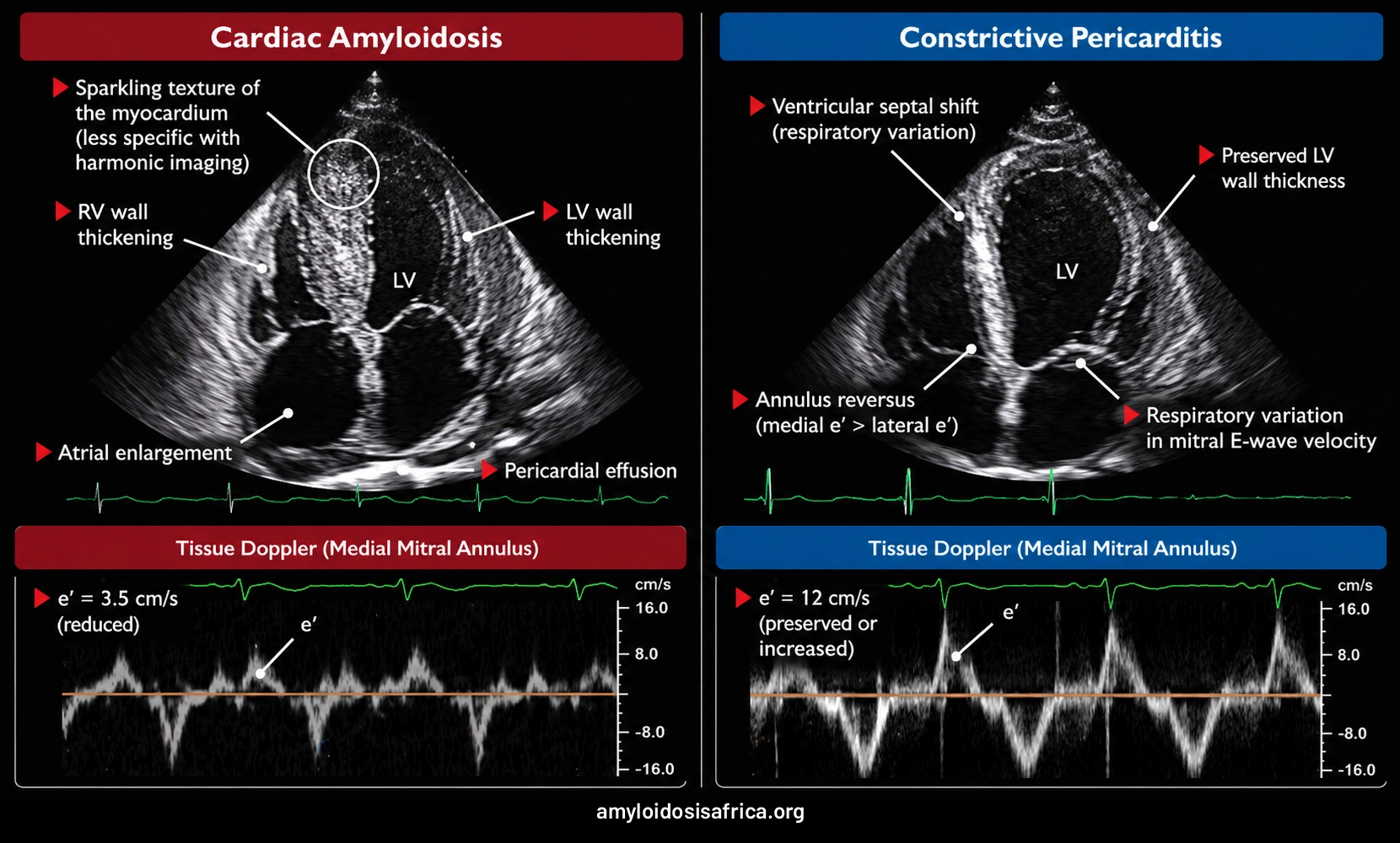
Figure 1. Echocardiographic Differences Between Cardiac Amyloidosis and Constrictive Pericarditis. Cardiac amyloidosis (left) is illustrated by increased ventricular wall thickness, atrial enlargement, and pericardial effusion, with reduced mitral annular early diastolic velocity (e′). Constrictive pericarditis (right) is associated with respiratory ventricular septal shift, preserved or increased medial e′ velocity, and possible annulus reversus (medial e′ greater than lateral e′). These findings help differentiate intrinsic myocardial restriction from pericardial constriction but require integrated clinical and echocardiographic assessment.
The Mayo Clinic Echocardiographic Criteria for Constrictive Pericarditis
The Mayo Clinic criteria provide a structured approach to differentiating constrictive pericarditis from restrictive myocardial disease.
Important findings include:
- respiratory ventricular septal shift;
- preserved or increased medial mitral annular e’; and
- prominent expiratory diastolic flow reversal in the hepatic veins.
The combination of ventricular septal shift with either medial e’ of at least 9 cm/s or a hepatic vein expiratory diastolic reversal ratio of at least 0.79 has demonstrated high diagnostic performance in the original study population.
However, these criteria are not infallible, and their interpretation may be affected by atrial fibrillation, loading conditions, mixed myocardial-pericardial disease and technical limitations.
Global Longitudinal Strain: Can GLS Help?
Global longitudinal strain measures longitudinal myocardial deformation and can provide additional information about myocardial function.
GLS in Cardiac Amyloidosis
Cardiac amyloidosis frequently produces reduced longitudinal strain, often with relatively preserved apical strain compared with basal and mid-ventricular segments.
This pattern is known as relative apical sparing.
However, relative apical sparing is not specific to amyloidosis and may be absent in early disease.
GLS in Constrictive Pericarditis
In constrictive pericarditis, longitudinal deformation may be relatively preserved compared with intrinsic restrictive myocardial disease, although abnormalities can occur.
Pericardial tethering and altered ventricular mechanics may affect regional strain patterns.
Therefore, GLS may support the distinction but cannot independently establish the diagnosis.
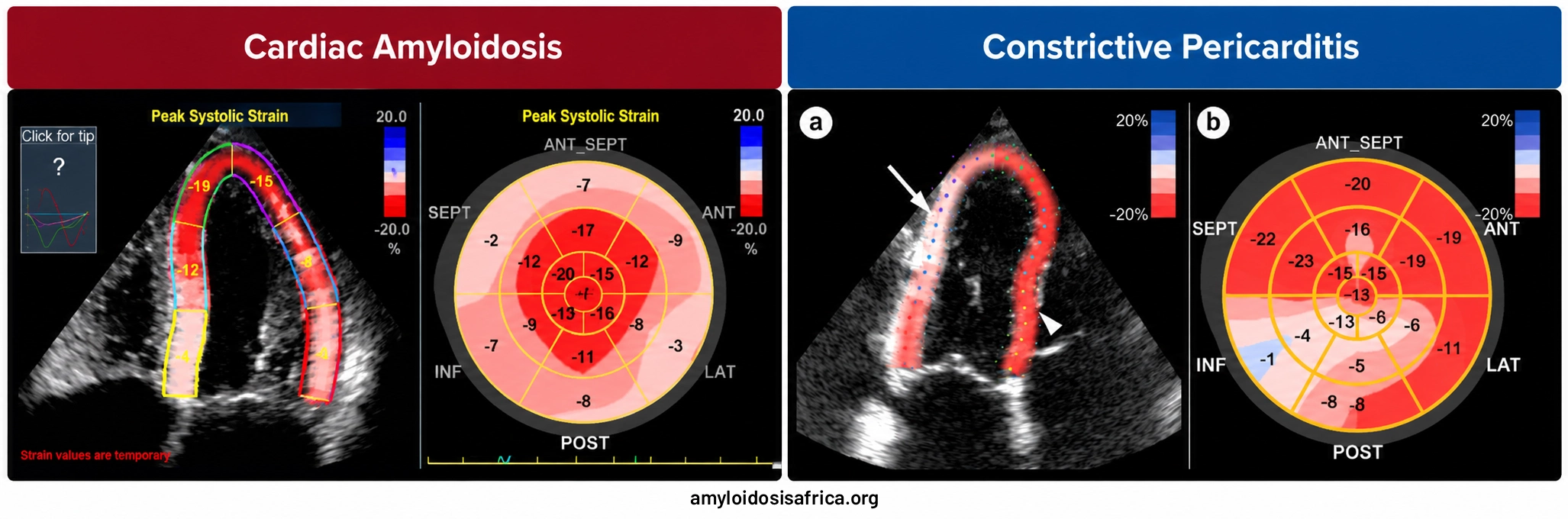
Figure 2. Global Longitudinal Strain in Cardiac Amyloidosis and Constrictive Pericarditis. Published speckle-tracking echocardiographic images illustrating differences in myocardial deformation. Cardiac amyloidosis (left) demonstrates relative apical sparing, with greater longitudinal strain impairment in the basal segments. Constrictive pericarditis (right) demonstrates a different regional strain distribution, with relatively greater impairment in selected inferolateral segments. These patterns may provide diagnostic clues but are not independently diagnostic and should be interpreted alongside Doppler echocardiography and other clinical findings.
Cardiac MRI: Myocardial Infiltration vs Pericardial Disease
Cardiac magnetic resonance imaging can be particularly helpful when echocardiographic findings are inconclusive.
CMR provides information about myocardial tissue characteristics, pericardial anatomy, inflammation, and ventricular interdependence.
CMR Findings in Cardiac Amyloidosis
Cardiac amyloidosis commonly demonstrates:
- diffuse subendocardial or transmural late gadolinium enhancement (LGE);
- elevated native T1 values;
- increased extracellular volume fraction (ECV);
- abnormal myocardial nulling; and
- increased ventricular wall thickness.
These findings reflect extracellular amyloid infiltration and associated myocardial tissue abnormalities.
However, imaging appearances vary with disease subtype and stage.
CMR Findings in Constrictive Pericarditis
CMR may demonstrate:
- pericardial thickening;
- pericardial late gadolinium enhancement;
- pericardial edema in inflammatory cases;
- abnormal septal motion; and
- respiratory ventricular interdependence on real-time cine imaging.
Importantly, a normal pericardial thickness does not exclude constrictive pericarditis.
Pericardial enhancement may suggest active inflammation and help identify patients who could benefit from anti-inflammatory treatment in appropriate circumstances.
Conversely, diffuse myocardial LGE with markedly increased ECV strongly favors an infiltrative myocardial process such as cardiac amyloidosis.
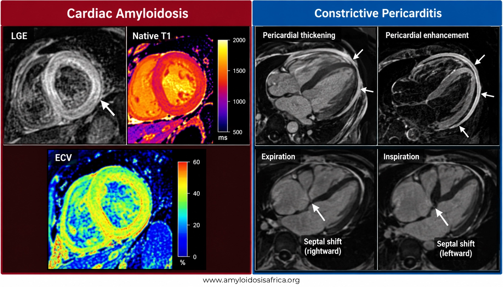
Figure 3. Cardiac MRI Differences Between Cardiac Amyloidosis and Constrictive Pericarditis. Illustrative cardiac magnetic resonance (CMR) comparison highlighting distinct myocardial and pericardial abnormalities. Cardiac amyloidosis (left) commonly demonstrates diffuse subendocardial or transmural late gadolinium enhancement (LGE), elevated native T1 and increased extracellular volume (ECV), reflecting myocardial amyloid infiltration. Constrictive pericarditis (right) may demonstrate pericardial thickening, pericardial enhancement in inflammatory disease and respiratory-dependent ventricular septal shift, reflecting exaggerated ventricular interdependence. These findings help differentiate intrinsic myocardial restriction from pericardial constriction and should be interpreted alongside clinical, echocardiographic, and hemodynamic assessments.
Publication note: This figure is an AI-generated educational illustration, not a collection of published patient MRI scans. It should be identified as illustrative and should not be attributed to clinical imaging publications.
Cardiac Catheterization: When Is Invasive Hemodynamic Assessment Needed?
Invasive hemodynamic assessment may be necessary when noninvasive investigations remain inconclusive or when mixed myocardial-pericardial disease is suspected.
Both cardiac amyloidosis and constrictive pericarditis may produce elevated ventricular filling pressures and a dip-and-plateau, or square-root, ventricular pressure contour.
Therefore, the presence of a square-root sign alone cannot distinguish the conditions.
Hemodynamics in Constrictive Pericarditis
A key finding is discordant respiratory variation in left and right ventricular systolic pressures.
During inspiration, right ventricular systolic pressure may increase while left ventricular systolic pressure decreases.
This reflects exaggerated ventricular interdependence.
Hemodynamics in Cardiac Amyloidosis
In myocardial restrictive physiology, left and right ventricular systolic pressures generally vary concordantly with respiration.
Elevated filling pressures reflect intrinsic myocardial stiffness rather than external pericardial restraint.
Simultaneous left and right ventricular pressure measurements may therefore help distinguish the two conditions.
Comparison Table: Cardiac Amyloidosis vs Constrictive Pericarditis
| Feature | Cardiac Amyloidosis | Constrictive Pericarditis |
|---|---|---|
| Primary abnormality | Myocardial amyloid infiltration | Noncompliant pericardium |
| Underlying mechanism | Intrinsic myocardial restriction | External restriction of cardiac filling |
| LV wall thickness | Frequently increased | Often normal |
| Septal bounce | Not characteristic | Common |
| Respiratory septal shift | Usually absent | Characteristic |
| Medial mitral e’ | Typically reduced | May be preserved or increased |
| Annulus reversus | Not typical | May be present |
| Hepatic vein expiratory reversal | Less characteristic | Supportive of constriction |
| GLS | Often reduced | May be relatively preserved |
| CMR myocardial LGE | Often diffuse subendocardial or transmural | Usually not an infiltrative pattern |
| Native T1 | Commonly elevated | May remain normal without myocardial disease |
| ECV | Frequently substantially increased | Not typically markedly expanded in isolated constriction |
| Pericardial thickening | Not a defining feature | May be present but is not required |
| Invasive hemodynamics | Respiratory ventricular systolic pressure concordance | Respiratory ventricular systolic pressure discordance |
| Treatment | Amyloid subtype-specific treatment | Anti-inflammatory therapy in selected cases or pericardiectomy |
Clinical Red Flags Suggesting Cardiac Amyloidosis
When restrictive physiology is identified, several clinical findings may increase suspicion for cardiac amyloidosis.
- bilateral carpal tunnel syndrome;
- peripheral or autonomic neuropathy;
- lumbar spinal stenosis;
- atraumatic biceps tendon rupture;
- unexplained increased ventricular wall thickness;
- conduction disease or atrial arrhythmias;
- disproportionately elevated cardiac biomarkers; and
- characteristic myocardial tissue abnormalities on CMR.
These manifestations vary according to amyloid subtype.
Macroglossia and periorbital purpura, when present, are particularly suggestive of AL amyloidosis.
Clinical Clues Suggesting Constrictive Pericarditis
Features that may increase suspicion for constrictive pericarditis include:
- previous tuberculosis or pericardial infection;
- prior cardiac surgery;
- history of chest radiation;
- previous recurrent pericarditis;
- prominent systemic venous congestion;
- Kussmaul sign;
- pericardial knock; and
- characteristic ventricular interdependence on echocardiography or CMR.
However, none of these findings alone establishes the diagnosis.
Diagnostic Approach to Unexplained Restrictive Physiology
Step 1: Assess Clinical History and Examination
Evaluate symptoms, previous pericardial disease, tuberculosis exposure, cardiac surgery, systemic inflammatory disease and extracardiac features suggestive of amyloidosis.
Step 2: Perform Comprehensive Echocardiography
Assess ventricular wall thickness, septal motion, tissue Doppler velocities, respiratory inflow variation and hepatic vein Doppler patterns.
Step 3: Consider Cardiac MRI
CMR can assess myocardial infiltration, pericardial thickness, inflammation and respiratory ventricular interdependence.
Step 4: Investigate Suspected Cardiac Amyloidosis
When cardiac amyloidosis is suspected, evaluate for AL amyloidosis using:
- serum free light chains with kappa/lambda ratio;
- serum immunofixation; and
- urine immunofixation.
These tests are essential because AL amyloidosis may require urgent hematological treatment.
When AL amyloidosis has been appropriately excluded, technetium-99m PYP, DPD or HMDP scintigraphy with SPECT can support nonbiopsy diagnosis of ATTR-CM in the appropriate clinical setting.
If a monoclonal protein is present or findings are discordant, tissue biopsy with accurate amyloid typing may be necessary.
Step 5: Consider Invasive Hemodynamic Assessment
When noninvasive findings are equivocal, simultaneous right and left ventricular catheterization may help differentiate constrictive from restrictive physiology.
Can Cardiac Amyloidosis and Constrictive Pericarditis Coexist?
Yes. Although uncommon, myocardial disease and pericardial constriction can coexist.
For example, patients with previous radiation exposure may develop both myocardial injury and pericardial disease.
Mixed physiology can complicate interpretation of echocardiographic and invasive hemodynamic findings.
Consequently, a diagnosis of constrictive pericarditis does not automatically exclude concurrent myocardial disease.
Similarly, identifying myocardial abnormalities does not completely exclude an additional pericardial component.
Why the Correct Diagnosis Changes Treatment
Treatment of Cardiac Amyloidosis
Management depends on the amyloid subtype.
ATTR-CM may be treated with disease-modifying therapies that target transthyretin, depending on clinical indication and local availability.
AL amyloidosis requires urgent treatment of the underlying plasma-cell disorder.
Management also includes appropriate treatment of heart failure, arrhythmias and conduction abnormalities.
Treatment of Constrictive Pericarditis
Management depends on the underlying cause, inflammatory activity and severity of constriction.
Patients with transient or inflammatory constriction may improve with appropriate anti-inflammatory treatment.
Chronic symptomatic constrictive pericarditis may require surgical pericardiectomy, particularly when medical management is insufficient.
However, surgical outcomes depend on patient selection, disease severity, etiology and the presence of concomitant myocardial dysfunction.
Accurate differentiation is therefore essential before considering surgery.
Why This Distinction Matters in Africa
Distinguishing cardiac amyloidosis from constrictive pericarditis is particularly relevant in African healthcare settings, where tuberculosis remains an important cause of pericardial disease in some populations.
At the same time, cardiac amyloidosis may be underrecognized because of limited access to specialized diagnostic investigations.
Both conditions may present with heart failure, ascites and systemic venous congestion, potentially leading to diagnostic uncertainty.
Challenges may include limited availability of:
- advanced echocardiography and tissue Doppler assessment;
- cardiac MRI;
- invasive hemodynamic testing;
- monoclonal protein testing;
- bone scintigraphy;
- specialized amyloid typing; and
- experienced multidisciplinary cardiomyopathy and pericardial disease teams.
Improving awareness of constrictive physiology and infiltrative cardiomyopathy may help clinicians identify patients who require specialized diagnostic evaluation.
Importantly, neither the presence of tuberculosis risk factors nor a history of pericarditis should automatically exclude other causes of restrictive physiology.
Key Takeaways
- Cardiac amyloidosis primarily affects the myocardium, while constrictive pericarditis primarily affects the pericardium.
- Both conditions can cause restrictive filling and right-sided heart failure symptoms.
- Respiratory septal shift and ventricular interdependence strongly support constrictive physiology.
- Preserved or increased medial mitral annular e’ favors constrictive pericarditis.
- Reduced annular velocities and increased ventricular wall thickness may support myocardial infiltration.
- Diffuse myocardial LGE, elevated native T1 and increased ECV favor cardiac amyloidosis.
- Pericardial thickening is supportive but not required for constrictive pericarditis.
- Invasive hemodynamic assessment may be useful when noninvasive findings remain inconclusive.
- Accurate diagnosis is essential because treatments differ substantially.
Frequently Asked Questions
Can cardiac amyloidosis mimic constrictive pericarditis?
Yes. Both conditions can cause elevated filling pressures, diastolic dysfunction, systemic venous congestion and heart failure with preserved ejection fraction.
What is the main difference between restrictive cardiomyopathy and constrictive pericarditis?
Restrictive cardiomyopathy results primarily from intrinsic myocardial dysfunction, whereas constrictive pericarditis results from external limitation of ventricular filling by a noncompliant pericardium.
What is septal bounce?
Septal bounce is abnormal interventricular septal motion commonly observed in constrictive pericarditis. It reflects altered ventricular interactions but is not independently diagnostic.
What is annulus reversus?
Annulus reversus occurs when medial mitral annular e’ exceeds lateral e’, reversing the usual relationship. It is a supportive echocardiographic finding in constrictive pericarditis.
Can constrictive pericarditis occur without pericardial thickening?
Yes. Constrictive physiology can occur even when pericardial thickness is within normal limits.
Does cardiac amyloidosis cause pericardial effusion?
Yes. Small pericardial effusions may occur in cardiac amyloidosis, but their presence does not establish constrictive pericarditis.
Can cardiac MRI distinguish the two conditions?
CMR can provide valuable evidence by identifying myocardial infiltration, pericardial abnormalities and ventricular interdependence. However, findings must be integrated with clinical and hemodynamic assessment.
Is the square-root sign specific for constrictive pericarditis?
No. A dip-and-plateau ventricular pressure contour can occur in both constrictive pericarditis and restrictive cardiomyopathy.
Can constrictive pericarditis be cured?
Some patients with inflammatory or transient constriction improve with medical treatment. In selected patients with chronic symptomatic constriction, pericardiectomy may provide substantial improvement.
References
- Welch TD, Ling LH, Espinosa RE, et al. Echocardiographic diagnosis of constrictive pericarditis: Mayo Clinic criteria. Circ Cardiovasc Imaging. 2014;7(3):526–534. doi:10.1161/CIRCIMAGING.113.001613.
- Garcia-Pavia P, Rapezzi C, Adler Y, et al. Diagnosis and treatment of cardiac amyloidosis: a position statement of the ESC Working Group on Myocardial and Pericardial Diseases. Eur Heart J. 2021;42(16):1554–1568. doi:10.1093/eurheartj/ehab072.
- Adler Y, Charron P, Imazio M, et al. 2015 ESC Guidelines for the diagnosis and management of pericardial diseases. Eur Heart J. 2015;36(42):2921–2964. doi:10.1093/eurheartj/ehv318.
- Gillmore JD, Maurer MS, Falk RH, et al. Nonbiopsy diagnosis of cardiac transthyretin amyloidosis. Circulation. 2016;133(24):2404–2412. doi:10.1161/CIRCULATIONAHA.116.021612.
- Kittleson MM, Ruberg FL, Ambardekar AV, et al. 2023 ACC Expert Consensus Decision Pathway on Comprehensive Multidisciplinary Care for the Patient With Cardiac Amyloidosis. J Am Coll Cardiol. 2023;81(11):1076–1126. doi:10.1016/j.jacc.2022.11.022.
This article is intended for educational purposes and does not replace individualized medical advice, specialist assessment or current clinical guidelines. Diagnosis and treatment decisions should be made by qualified healthcare professionals based on the patient’s clinical findings and appropriate investigations.

Leave a Reply