



Cardiac Amyloidosis vs Hypertensive Heart Disease: How to Tell Them Apart
Cardiac amyloidosis vs hypertensive heart disease is an important diagnostic comparison because both conditions can cause increased left ventricular wall thickness, diastolic dysfunction, left atrial enlargement and heart failure with preserved ejection fraction (HFpEF).
In clinical practice, physicians often attribute ventricular thickening to longstanding hypertension. Although this explanation may be correct, it can sometimes delay recognition of cardiac amyloidosis, particularly transthyretin amyloid cardiomyopathy (ATTR-CM) in older adults.
The distinction matters because hypertension-related myocardial remodeling and amyloid infiltration involve different pathological processes. Furthermore, their management strategies differ substantially.
Importantly, hypertension and cardiac amyloidosis can coexist. Therefore, clinicians should not exclude amyloidosis simply because a patient has a history of elevated blood pressure.
This article explains how clinical findings, electrocardiography (ECG), echocardiography, global longitudinal strain (GLS), cardiac magnetic resonance imaging (CMR), and disease-specific investigations help distinguish these conditions (1, 2).
What Is Hypertensive Heart Disease?
Hypertensive heart disease refers to structural and functional cardiac changes associated with chronic elevation of systemic arterial blood pressure.
Persistent pressure overload increases left ventricular wall stress. In response, cardiomyocytes enlarge, and the myocardium may develop interstitial fibrosis and impaired relaxation.
Over time, these changes can lead to:
- concentric left ventricular hypertrophy (LVH);
- increased left ventricular mass;
- impaired ventricular relaxation and diastolic dysfunction;
- left atrial enlargement;
- reduced longitudinal myocardial function;
- atrial fibrillation; and
- heart failure, with preserved or reduced ejection fraction.
However, the severity of myocardial remodeling varies according to blood pressure burden, age, genetic factors, obesity, kidney disease, and other comorbidities.
Consequently, not every patient with hypertension develops significant LVH, and the presence of LVH does not automatically establish hypertension as its cause.
What Is Cardiac Amyloidosis?
Cardiac amyloidosis develops when abnormal protein fibrils accumulate in the myocardial extracellular space. These deposits increase ventricular wall thickness and progressively impair myocardial relaxation and function.
The two major forms are:
- Transthyretin amyloidosis (ATTR): caused by misfolded transthyretin, either in hereditary ATTR or wild-type ATTR.
- Immunoglobulin light-chain amyloidosis (AL): caused by amyloidogenic light chains associated with an underlying clonal plasma-cell disorder.
Unlike hypertensive remodeling, amyloid infiltration increases apparent wall thickness through extracellular deposition rather than cardiomyocyte hypertrophy alone.
As the disease progresses, patients may develop restrictive ventricular filling, reduced stroke volume, atrial arrhythmias, conduction disease, and heart failure.
Learn more about wild-type ATTR amyloidosis, hereditary ATTR amyloidosis and AL amyloidosis.
Why Do These Conditions Look Similar?
Both diseases can increase ventricular wall thickness while preserving left ventricular ejection fraction during earlier stages.
In addition, both may produce diastolic dysfunction, atrial enlargement, impaired longitudinal strain, and symptoms of heart failure.
However, the underlying mechanisms differ:
- Hypertensive heart disease: chronic pressure overload promotes cardiomyocyte hypertrophy and variable myocardial fibrosis.
- Cardiac amyloidosis: extracellular amyloid deposits expand the interstitial compartment and impair myocardial mechanical function.
These differences become particularly relevant when clinicians evaluate ventricular geometry, tissue characterization and extracardiac manifestations.
Ventricular Hypertrophy: Can Wall Thickness Distinguish Them?
Hypertensive Heart Disease
Hypertension commonly produces concentric remodeling or concentric hypertrophy, characterized by increased relative wall thickness and, in established LVH, increased left ventricular mass.
Nevertheless, some patients develop eccentric remodeling or other geometric patterns, especially when additional cardiac conditions coexist.
The degree of hypertrophy generally reflects cumulative pressure exposure and individual susceptibility. However, blood pressure measurements at a single visit may not represent the patient’s historical blood pressure burden.
Cardiac Amyloidosis
Cardiac amyloidosis also frequently produces a concentric pattern of increased wall thickness.
Additional findings may include thickening of the right ventricular free wall, interatrial septum, and atrioventricular valves.
Importantly, amyloidosis may produce asymmetric wall thickening, particularly in ATTR-CM. Therefore, neither concentric nor asymmetric morphology is independently diagnostic.
According to the European Society of Cardiology position statement, increased left ventricular wall thickness of at least 12 mm accompanied by suggestive clinical or imaging findings should prompt consideration of cardiac amyloidosis (1).
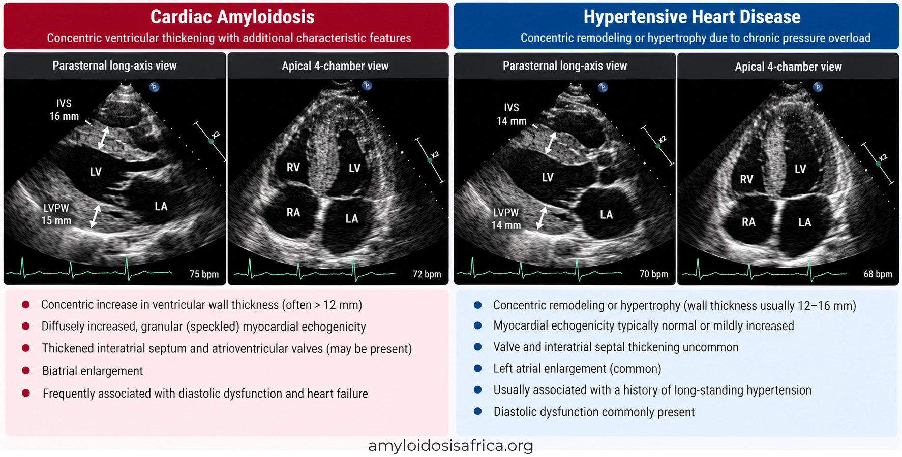
Figure 1. Ventricular Wall Thickening in Cardiac Amyloidosis and Hypertensive Heart Disease. Both conditions can produce concentric left ventricular hypertrophy and diastolic dysfunction. Cardiac amyloidosis is characterized by extracellular amyloid deposition and may be associated with biatrial enlargement, interatrial septal thickening, and atrioventricular valve thickening. In contrast, hypertensive heart disease primarily reflects chronic pressure-overload remodeling, often accompanied by left atrial enlargement. These findings may overlap, and echocardiography alone cannot reliably distinguish the two conditions.
Echocardiography: Important Diagnostic Clues
Echocardiography is usually the first-line imaging investigation when clinicians identify increased ventricular wall thickness.
However, accurate interpretation requires more than measuring the interventricular septum and posterior wall.
Clinicians should assess ventricular geometry, systolic and diastolic function, atrial dimensions, valve appearance, right ventricular involvement, and myocardial deformation.
| Feature | Cardiac Amyloidosis | Hypertensive Heart Disease |
|---|---|---|
| LV wall thickness | Often diffuse or concentric; may be asymmetric | Often concentric remodeling or hypertrophy |
| LV mass | Frequently increased with infiltration | May increase with chronic pressure overload |
| Diastolic dysfunction | Common; may progress to restrictive filling | Common; severity varies |
| Left atrial enlargement | Common | Common |
| Right ventricular wall thickening | May provide an additional clue | Less characteristic in uncomplicated systemic hypertension |
| Valve or interatrial septal thickening | May accompany amyloid infiltration | Not a characteristic consequence of hypertension alone |
| GLS | Often reduced with relative apical sparing | May be reduced, particularly in more affected segments |
| Ejection fraction | Often preserved initially | May remain preserved for many years |
Although these findings are useful, no single echocardiographic feature can definitively distinguish cardiac amyloidosis from hypertensive heart disease.
Global Longitudinal Strain: The Role of Apical Sparing
Global longitudinal strain measures longitudinal myocardial deformation using speckle-tracking echocardiography.
GLS can identify impaired myocardial function even when left ventricular ejection fraction remains normal.
Strain in Cardiac Amyloidosis
Cardiac amyloidosis frequently causes greater longitudinal strain impairment in basal and mid-ventricular segments than at the apex.
This creates the characteristic relative apical-sparing pattern on a GLS bull’s-eye map.
However, relative apical sparing is not specific to amyloidosis. It can occur in other cardiac conditions, and some patients with amyloidosis do not demonstrate a classic pattern.
Strain in Hypertensive Heart Disease
Hypertensive heart disease can also reduce GLS, sometimes before conventional ejection fraction measurements become abnormal.
Longitudinal dysfunction may involve basal or other myocardial segments, depending on disease severity and associated fibrosis.
Consequently, a relative base-to-apex strain gradient does not automatically establish cardiac amyloidosis.
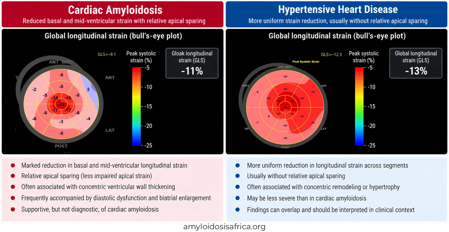
Figure 2. Global Longitudinal Strain in Cardiac Amyloidosis and Hypertensive Heart Disease. Cardiac amyloidosis frequently demonstrates reduced basal and mid-ventricular longitudinal strain with relative preservation of apical strain (apical sparing). In contrast, hypertensive heart disease may produce more uniform or heterogeneous longitudinal strain impairment associated with pressure-overload remodeling and myocardial fibrosis. However, these patterns can overlap, and relative apical sparing is not specific to cardiac amyloidosis. GLS findings should be interpreted alongside clinical assessment and other diagnostic investigations.
ECG Findings: Can Low Voltage Help?
Electrocardiography provides additional information when clinicians evaluate ventricular hypertrophy.
Hypertensive heart disease may produce increased QRS voltage, repolarization abnormalities, and left ventricular hypertrophy patterns.
In contrast, cardiac amyloidosis can cause relatively low QRS voltage despite increased ventricular wall thickness on echocardiography.
This discrepancy between electrical voltage and anatomical wall thickness is a recognized amyloidosis clue.
However, low voltage is absent in many patients with ATTR-CM. Moreover, obesity, pulmonary disease, and other conditions can affect ECG voltage.
Therefore, low voltage supports suspicion in the appropriate setting but does not confirm amyloidosis, while normal or high voltage does not exclude it (2).
Cardiac MRI: Late Gadolinium Enhancement
Cardiac magnetic resonance imaging provides detailed assessment of myocardial morphology, tissue characteristics, and fibrosis.
It becomes particularly useful when echocardiography cannot adequately explain increased ventricular wall thickness.
LGE in Cardiac Amyloidosis
Cardiac amyloidosis often produces diffuse subendocardial or transmural late gadolinium enhancement (LGE).
Additional features may include abnormal myocardial nulling and extensive extracellular expansion.
These findings reflect amyloid deposition and altered myocardial tissue composition.
LGE in Hypertensive Heart Disease
Hypertensive heart disease may demonstrate no LGE or focal nonischemic enhancement, often in a mid-wall distribution.
When present, enhancement generally reflects replacement fibrosis associated with chronic myocardial stress.
However, LGE patterns vary, particularly when coronary artery disease or another cardiomyopathy coexists.
Therefore, clinicians should interpret enhancement patterns together with native T1 mapping, extracellular volume, and the complete clinical phenotype.
Native T1 Mapping and Extracellular Volume
Native T1 Mapping
Native T1 mapping measures myocardial tissue characteristics without requiring gadolinium contrast.
Cardiac amyloidosis frequently produces elevated native T1 values because of myocardial infiltration and extracellular expansion.
Hypertensive heart disease may also increase native T1, particularly when diffuse myocardial fibrosis develops. However, values are often less markedly abnormal than in advanced cardiac amyloidosis.
Importantly, native T1 values vary according to scanner field strength, imaging sequence, and local reference ranges. Therefore, no universal cutoff reliably separates these diseases.
Extracellular Volume
Extracellular volume fraction (ECV) estimates the proportion of myocardial tissue occupied by the extracellular compartment.
Cardiac amyloidosis frequently causes substantial ECV expansion because amyloid fibrils accumulate outside cardiomyocytes.
Hypertensive heart disease can also increase ECV through interstitial fibrosis, but the expansion is generally less extensive than in advanced amyloid infiltration.
Nevertheless, ECV values overlap, particularly in earlier disease stages. Thus, ECV is an important supportive measurement rather than an independent confirmatory test.
| CMR Feature | Cardiac Amyloidosis | Hypertensive Heart Disease |
|---|---|---|
| LGE | Often diffuse subendocardial or transmural | May be absent or show focal mid-wall fibrosis |
| Native T1 | Frequently markedly elevated | Normal or elevated depending on fibrosis |
| ECV | Often substantially increased | May be moderately increased |
| Myocardial nulling | May show characteristic abnormalities | Usually lacks the typical amyloid nulling pattern |
| Underlying tissue process | Extracellular amyloid deposition | Myocyte hypertrophy and myocardial fibrosis |
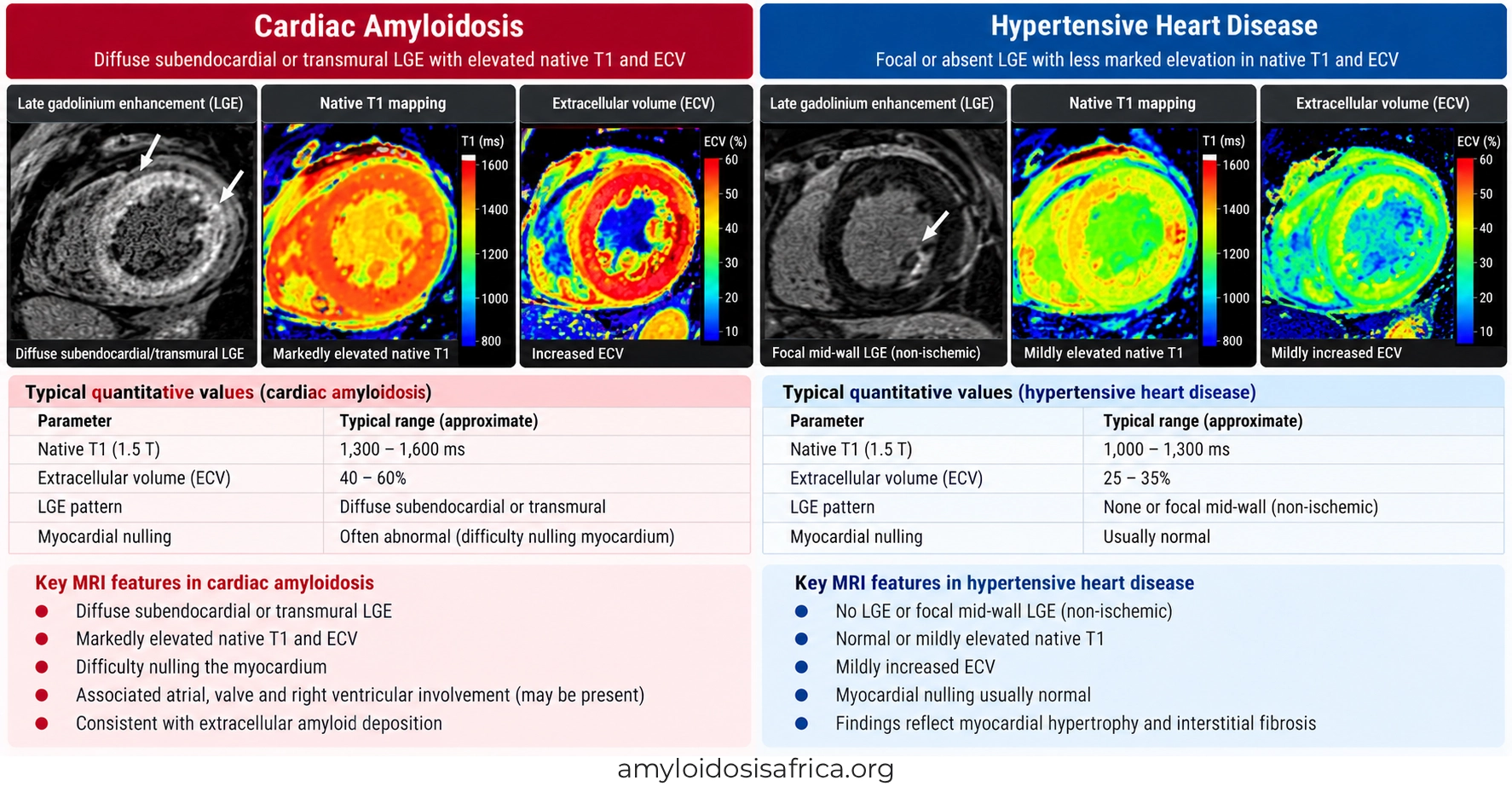
Figure 3. Cardiac MRI Differences Between Cardiac Amyloidosis and Hypertensive Heart Disease. Cardiac amyloidosis commonly demonstrates diffuse subendocardial or transmural late gadolinium enhancement (LGE), elevated native T1 values, and substantial extracellular volume (ECV) expansion, reflecting myocardial amyloid deposition. In contrast, hypertensive heart disease may show absent or focal mid-wall LGE, with less pronounced increases in native T1 and ECV associated with myocardial hypertrophy and interstitial fibrosis. These imaging characteristics provide important diagnostic clues, although findings may overlap and should be interpreted alongside clinical assessment and other investigations.
Clinical Red Flags That Favor Cardiac Amyloidosis
Clinical findings outside the heart can provide important diagnostic clues.
Features that should increase suspicion for cardiac amyloidosis include:
- heart failure with preserved ejection fraction and unexplained ventricular thickening;
- bilateral carpal tunnel syndrome;
- lumbar spinal stenosis;
- atraumatic biceps tendon rupture;
- peripheral neuropathy;
- autonomic dysfunction or orthostatic hypotension;
- conduction disease or pacemaker implantation;
- unexplained proteinuria;
- macroglossia or periorbital purpura, particularly in AL amyloidosis; and
- persistently elevated cardiac biomarkers without another adequate explanation.
These findings become particularly important when cardiac imaging appears disproportionate to the documented blood pressure history.
For example, an older patient with controlled hypertension, progressive heart failure, bilateral carpal tunnel syndrome and increased ventricular wall thickness should undergo further assessment for possible ATTR-CM.
Can Hypertension and Cardiac Amyloidosis Coexist?
Yes. Hypertension is common in the general population, particularly among older adults, and may coexist with ATTR or AL cardiac amyloidosis.
Therefore, clinicians should not treat these diagnoses as mutually exclusive.
In some patients, longstanding hypertension contributes to ventricular remodeling before amyloid infiltration becomes clinically apparent.
Additionally, progressive amyloid cardiomyopathy may reduce cardiac output and blood pressure, causing patients who previously required several antihypertensive medications to develop lower blood pressure or treatment intolerance.
However, falling blood pressure is nonspecific and may reflect medication effects, dehydration, autonomic dysfunction, or other illnesses.
Consequently, it should be interpreted as one potential clue rather than a diagnostic criterion.
When Should Clinicians Suspect Amyloidosis Despite Hypertension?
Further investigation becomes particularly important when:
- ventricular wall thickening appears disproportionate to the severity or duration of hypertension;
- heart failure progresses despite apparently adequate blood pressure management;
- ECG voltage appears unexpectedly low relative to wall thickness;
- echocardiography shows relative apical sparing alongside other suspicious features;
- CMR demonstrates diffuse subendocardial or transmural LGE;
- neurologic, orthopedic or autonomic red flags are present; or
- conduction abnormalities develop without another clear explanation.
These findings do not independently diagnose amyloidosis. Instead, they identify patients who may benefit from targeted testing.
Diagnostic Approach: How to Confirm the Correct Disease
Step 1: Review the Hypertension History
Assess previous blood pressure measurements, treatment duration, medication adherence, evidence of longstanding pressure overload and associated conditions.
Ambulatory or home blood pressure monitoring may help clarify the current blood pressure burden when clinically indicated.
Step 2: Evaluate ECG and Echocardiography
Review ventricular geometry, diastolic function, GLS, atrial size, right ventricular involvement, and electrical abnormalities.
Importantly, identify any mismatch between ventricular thickness and ECG voltage.
Step 3: Screen for AL Amyloidosis
When cardiac amyloidosis is suspected, obtain a complete monoclonal protein screen:
- serum free light chains, including the kappa/lambda ratio;
- serum immunofixation; and
- urine immunofixation.
These tests are essential because AL amyloidosis requires prompt specialist evaluation.
Serum or urine protein electrophoresis alone is insufficient to exclude AL amyloidosis (2).
Read more about monoclonal protein testing in cardiac amyloidosis.
Step 4: Consider Cardiac MRI
CMR may help identify characteristic myocardial infiltration and distinguish it from pressure-overload remodeling.
However, CMR cannot independently establish whether cardiac amyloid deposits consist of ATTR or AL proteins.
Learn more about cardiac MRI in cardiac amyloidosis.
Step 5: Investigate ATTR Cardiomyopathy
When monoclonal protein testing is negative, appropriate bone-tracer scintigraphy may support a nonbiopsy diagnosis of ATTR-CM.
In the correct clinical setting, grade 2 or 3 myocardial uptake on technetium-99m PYP, DPD, or HMDP scintigraphy, with SPECT confirmation and no evidence of a monoclonal protein, can establish ATTR-CM noninvasively (3).
However, when monoclonal testing is abnormal, scintigraphy alone cannot reliably distinguish ATTR from AL amyloidosis.
In such cases, tissue biopsy with accurate amyloid typing may be required.
Read more about bone scintigraphy and cardiac amyloidosis biopsy.
Step 6: Perform Genetic Testing When ATTR Is Confirmed
Once clinicians establish ATTR amyloidosis, TTR genetic testing distinguishes hereditary from wild-type disease.
This distinction informs genetic counseling, family assessment and appropriate long-term management.
Why the Correct Diagnosis Changes Treatment
Hypertensive Heart Disease
Management focuses on effective blood pressure control, cardiovascular risk reduction and treatment of associated heart failure, kidney disease and other comorbidities.
Depending on the clinical situation, clinicians may use antihypertensive agents, lifestyle interventions and guideline-directed heart failure therapies.
Effective blood pressure control can reduce cardiovascular risk and may lead to regression of hypertension-related LVH in some patients.
Cardiac Amyloidosis
Cardiac amyloidosis requires subtype-specific management.
ATTR-CM treatment may include transthyretin stabilizers or TTR-directed gene-silencing therapy according to regulatory approval, clinical indication and patient characteristics.
In contrast, AL amyloidosis requires treatment of the underlying clonal plasma-cell disorder.
Heart failure management also differs because patients with cardiac amyloidosis may have reduced tolerance of certain medications, particularly in advanced restrictive disease or autonomic dysfunction.
Therefore, incorrectly attributing amyloid cardiomyopathy to hypertension can delay appropriate treatment.
For a broader discussion, see cardiac amyloidosis treatment and heart failure management.
Summary: Cardiac Amyloidosis vs Hypertensive Heart Disease
| Diagnostic Feature | Cardiac Amyloidosis | Hypertensive Heart Disease |
|---|---|---|
| Underlying mechanism | Extracellular amyloid deposition | Chronic pressure-overload remodeling |
| Typical LV geometry | Often concentric thickening | Often concentric remodeling or LVH |
| ECG voltage | May be low relative to wall thickness | May demonstrate LVH voltage criteria |
| GLS | Relative apical sparing frequently observed | Reduced GLS possible; regional overlap occurs |
| CMR LGE | Often diffuse subendocardial or transmural | May be absent or show focal mid-wall fibrosis |
| Native T1 and ECV | Frequently markedly elevated | May increase with myocardial fibrosis |
| Extracardiac red flags | Carpal tunnel, neuropathy, spinal stenosis and others | Not characteristic of hypertension itself |
| Confirmatory testing | Monoclonal screen, scintigraphy, biopsy when indicated | Clinical blood pressure assessment and evaluation of other causes |
| Management | Amyloid subtype-specific treatment | Blood pressure and cardiovascular risk management |
Why This Distinction Matters in Africa
Hypertension is a major contributor to cardiovascular disease across Africa. Consequently, clinicians frequently encounter patients with increased ventricular wall thickness and heart failure in the setting of elevated blood pressure.
However, the presence of hypertension may create diagnostic anchoring, particularly when access to advanced cardiac imaging, immunofixation, serum free light chain assays, or bone-tracer scintigraphy is limited.
Cardiac amyloidosis should therefore remain part of the differential diagnosis when clinical and imaging findings appear atypical for hypertension alone.
Improving recognition requires stronger clinical education, appropriate diagnostic referral pathways, and expanded access to amyloidosis-specific testing.
Multicenter African research can also help clarify how often ATTR-CM or AL amyloidosis is missed among patients initially classified as having hypertensive heart disease.
Learn more about cardiac amyloidosis in Africa.
Key Takeaways
- Cardiac amyloidosis and hypertensive heart disease can both produce concentric ventricular thickening, diastolic dysfunction, and HFpEF.
- Hypertension causes pressure-overload remodeling, while cardiac amyloidosis causes extracellular protein deposition.
- A history of hypertension does not exclude cardiac amyloidosis.
- Relative apical sparing on GLS increases suspicion for amyloidosis but is not diagnostic.
- CMR LGE, native T1 and ECV mapping can help distinguish infiltrative disease from pressure-overload remodeling.
- Low ECG voltage relative to increased wall thickness is a useful clue but is not present in every patient.
- Extracardiac red flags can substantially strengthen suspicion for cardiac amyloidosis.
- When amyloidosis is suspected, a complete monoclonal protein screen is essential.
- Accurate diagnosis enables appropriate disease-specific treatment.
Frequently Asked Questions
Can hypertension cause the same wall thickening as cardiac amyloidosis?
Yes. Both conditions can increase left ventricular wall thickness, particularly in a concentric pattern. However, the underlying mechanisms differ, and further evaluation may be necessary when additional suspicious findings are present.
Can a patient have both hypertension and cardiac amyloidosis?
Yes. Hypertension and cardiac amyloidosis can coexist, especially in older adults. Therefore, hypertension should not automatically exclude amyloidosis from the differential diagnosis.
Does low ECG voltage confirm cardiac amyloidosis?
No. Low voltage relative to ventricular wall thickness is a recognized clue, but many patients with ATTR-CM do not have low QRS voltage.
Is apical sparing specific to cardiac amyloidosis?
No. Relative apical sparing can occur in other cardiac conditions, including hypertensive heart disease. It should be interpreted together with clinical findings and other investigations.
Which CMR findings favor cardiac amyloidosis?
Diffuse subendocardial or transmural LGE, elevated native T1, substantial ECV expansion and abnormal myocardial nulling support suspicion for cardiac amyloidosis.
Can hypertensive heart disease increase extracellular volume?
Yes. Myocardial fibrosis associated with hypertension can increase ECV. However, marked diffuse ECV expansion is more characteristic of advanced cardiac amyloidosis.
When should clinicians investigate cardiac amyloidosis in a patient with hypertension?
Further evaluation is appropriate when ventricular thickening is disproportionate to the hypertension history, unexplained heart failure develops, or ECG, strain, CMR, or extracardiac findings raise suspicion for amyloidosis.
Can cardiac MRI distinguish AL from ATTR amyloidosis?
No. CMR can support the diagnosis of cardiac amyloid infiltration but cannot reliably establish the amyloid subtype. Disease-specific laboratory testing, scintigraphy or biopsy may be necessary.
Why does early diagnosis matter?
Early recognition helps clinicians identify the correct disease mechanism, initiate appropriate treatment and avoid delays in managing potentially progressive cardiac amyloidosis.
References
- Garcia-Pavia P, Rapezzi C, Adler Y, et al. Diagnosis and treatment of cardiac amyloidosis: a position statement of the ESC Working Group on Myocardial and Pericardial Diseases. Eur Heart J. 2021;42(16):1554–1568. doi:10.1093/eurheartj/ehab072.
- Kittleson MM, Ruberg FL, Ambardekar AV, et al. 2023 ACC Expert Consensus Decision Pathway on Comprehensive Multidisciplinary Care for the Patient With Cardiac Amyloidosis. J Am Coll Cardiol. 2023;81(11):1076–1126. doi:10.1016/j.jacc.2022.11.022.
- Gillmore JD, Maurer MS, Falk RH, et al. Nonbiopsy Diagnosis of Cardiac Transthyretin Amyloidosis. Circulation. 2016;133(24):2404–2412. doi:10.1161/CIRCULATIONAHA.116.021612.
- Phelan D, Collier P, Thavendiranathan P, et al. Relative apical sparing of longitudinal strain using two-dimensional speckle-tracking echocardiography is both sensitive and specific for the diagnosis of cardiac amyloidosis. Heart. 2012;98(19):1442–1448. doi:10.1136/heartjnl-2012-302353.
This article provides educational information for healthcare professionals and the public. It does not replace individualized medical assessment, diagnostic investigations, or specialist recommendations. Cardiac amyloidosis and hypertensive heart disease may coexist and have overlapping clinical and imaging features. Diagnostic and treatment decisions should follow current professional guidelines and local regulatory requirements.



Leave a Reply