



Cardiac Amyloidosis vs Hypertrophic Cardiomyopathy: How to Tell Them Apart
Cardiac amyloidosis vs hypertrophic cardiomyopathy is an important diagnostic comparison because both conditions can cause increased left ventricular wall thickness, diastolic dysfunction, abnormal myocardial strain, and heart failure. Although their cardiac imaging findings sometimes overlap, the underlying diseases differ substantially.
Cardiac amyloidosis develops when misfolded proteins accumulate in the myocardial extracellular space. In contrast, hypertrophic cardiomyopathy (HCM) typically involves a primary myocardial disorder, often associated with pathogenic variants in genes encoding sarcomeric proteins.
These differences influence clinical presentation, diagnostic investigations, treatment, and prognosis. Therefore, identifying the correct cause of ventricular thickening is essential rather than relying on wall thickness alone.
This article explains how echocardiography, global longitudinal strain (GLS), cardiac magnetic resonance imaging (CMR), clinical red flags and targeted investigations help distinguish cardiac amyloidosis from HCM (1, 2).
What Is Cardiac Amyloidosis?
Cardiac amyloidosis is an infiltrative cardiomyopathy in which abnormal protein fibrils accumulate within the myocardial extracellular space. As amyloid deposits expand, ventricular walls become thicker and stiffer, impairing relaxation and filling.
The two major forms of cardiac amyloidosis are:
- Transthyretin amyloidosis (ATTR): caused by misfolding of transthyretin, either in hereditary ATTR or wild-type ATTR.
- Immunoglobulin light-chain amyloidosis (AL): caused by amyloidogenic immunoglobulin light chains produced by a clonal plasma-cell disorder.
Both types can cause increased ventricular wall thickness and restrictive cardiac physiology. However, their treatments differ considerably.
Learn more about wild-type ATTR amyloidosis, hereditary ATTR amyloidosis and AL amyloidosis.
What Is Hypertrophic Cardiomyopathy?
Hypertrophic cardiomyopathy is a myocardial disease characterized by increased ventricular wall thickness that cannot be explained solely by abnormal loading conditions, such as hypertension or aortic stenosis.
Many patients have pathogenic variants in sarcomeric genes, including MYH7 and MYBPC3. Nevertheless, genetic testing does not identify a causative variant in every patient with clinically diagnosed HCM.
HCM commonly produces asymmetric septal hypertrophy. However, other patterns include concentric, apical, and focal hypertrophy.
Some patients develop dynamic left ventricular outflow tract obstruction (LVOTO), which may contribute to exertional dyspnea, chest discomfort, and syncope.
The 2024 AHA/ACC multisociety guideline emphasizes comprehensive clinical evaluation, echocardiography, family history, and selective CMR to establish the diagnosis and distinguish HCM from alternative causes of ventricular hypertrophy (2).
Why Can These Two Diseases Look Similar?
Both conditions can produce increased ventricular wall thickness, small ventricular cavities, preserved ejection fraction, and impaired diastolic function.
Furthermore, patients may report similar symptoms, including exertional breathlessness, fatigue, chest discomfort, and reduced exercise capacity.
However, the mechanism of wall thickening differs.
- Cardiac amyloidosis: extracellular amyloid deposition expands the myocardial interstitial space.
- HCM: cardiomyocyte hypertrophy, myocyte disarray, and variable interstitial or replacement fibrosis contribute to increased myocardial mass.
Consequently, the combination of ventricular morphology, myocardial deformation and tissue characterization often provides more diagnostic information than wall thickness alone.
Ventricular Hypertrophy: Asymmetric vs Concentric Patterns
Typical Findings in HCM
HCM frequently produces asymmetric septal hypertrophy, with disproportionate thickening of the interventricular septum compared with other ventricular segments.
Additional findings may include:
- localized or apical hypertrophy;
- small left ventricular cavity;
- systolic anterior motion of the mitral valve;
- dynamic LV outflow tract obstruction;
- mitral regurgitation; and
- left atrial enlargement.
In adults, unexplained maximal left ventricular wall thickness of at least 15 mm generally supports a clinical diagnosis of HCM. A threshold of 13–14 mm may be relevant in selected patients, particularly those with a family history or other supportive evidence (2).
Typical Findings in Cardiac Amyloidosis
Cardiac amyloidosis commonly produces increased wall thickness involving multiple ventricular segments. A concentric appearance is frequent, particularly in advanced disease.
Additional features may include:
- biatrial enlargement;
- increased right ventricular wall thickness;
- thickening of atrioventricular valves;
- diastolic dysfunction;
- reduced longitudinal myocardial function;
- small pericardial effusion; and
- restrictive filling physiology in advanced disease.
Nevertheless, ATTR cardiomyopathy can produce asymmetric septal hypertrophy and may closely resemble HCM. Therefore, asymmetric morphology should not end the diagnostic evaluation when other findings suggest amyloidosis.
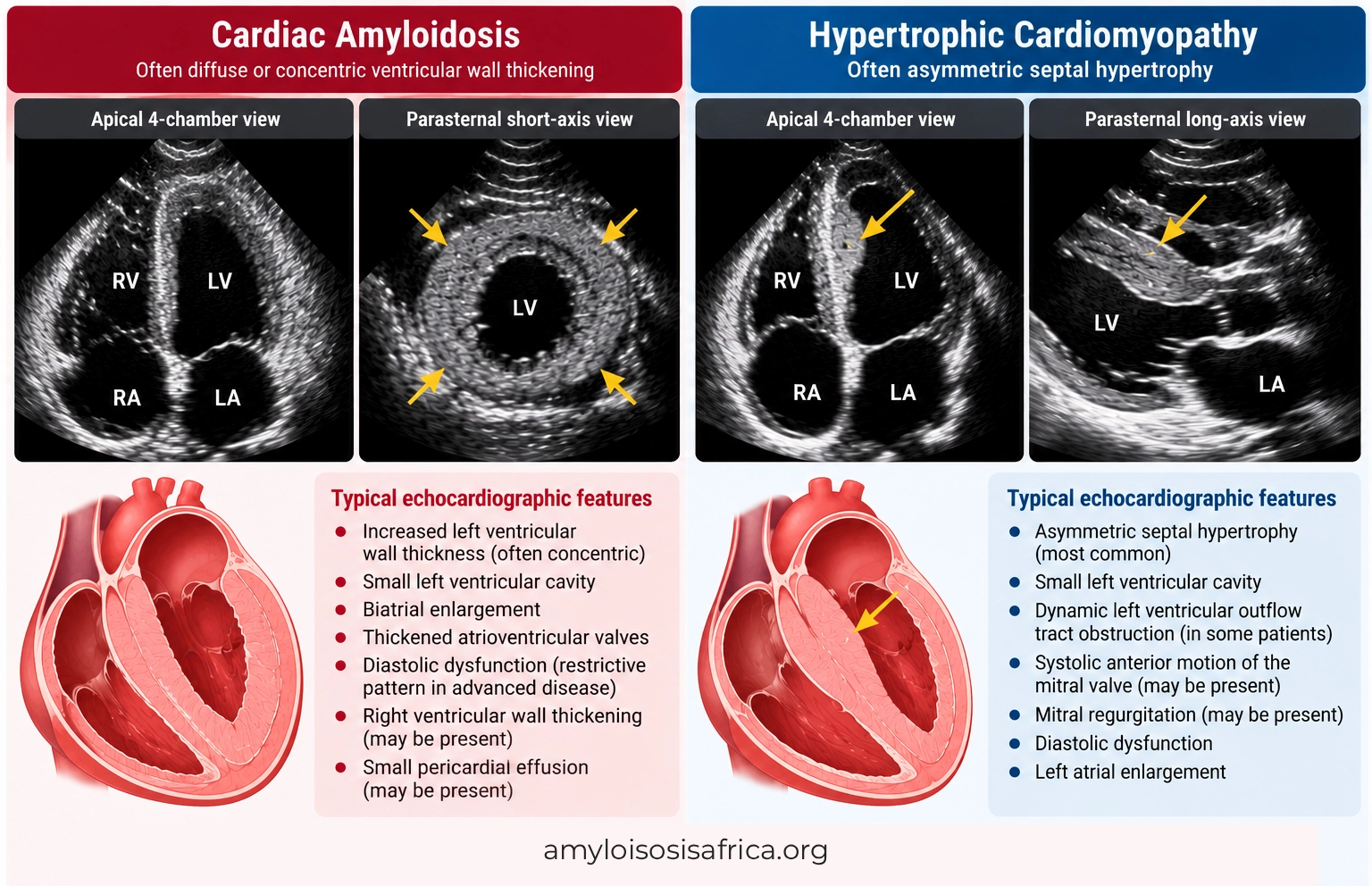
Figure 1. Ventricular Hypertrophy in Cardiac Amyloidosis and Hypertrophic Cardiomyopathy. Cardiac amyloidosis commonly produces diffuse or concentric ventricular wall thickening, often accompanied by diastolic dysfunction, biatrial enlargement, and other features of myocardial infiltration. In contrast, hypertrophic cardiomyopathy (HCM) frequently presents with asymmetric septal hypertrophy and may cause dynamic left ventricular outflow tract obstruction. However, these morphological patterns can overlap, and echocardiography alone cannot reliably distinguish the two conditions.
Echocardiography: Key Diagnostic Differences
Transthoracic echocardiography is usually the first-line imaging investigation when clinicians identify unexplained ventricular hypertrophy.
Beyond wall thickness, clinicians should evaluate ventricular geometry, diastolic function, mitral valve anatomy, outflow gradients, atrial size, and myocardial strain.
| Feature | Cardiac Amyloidosis | Hypertrophic Cardiomyopathy |
|---|---|---|
| LV wall thickness | Often diffuse or concentric; asymmetric patterns possible | Often asymmetric; concentric, apical and focal patterns possible |
| LV outflow obstruction | Less typical, but can occur | Common in obstructive HCM |
| Diastolic dysfunction | Common and may become restrictive | Common; severity varies |
| Left atrial enlargement | Common | Common |
| Valve thickening | May accompany myocardial infiltration | Mitral valve abnormalities may contribute to obstruction |
| GLS | Frequently reduced with relative apical sparing | Frequently reduced in hypertrophied or fibrotic segments |
| Ejection fraction | May remain preserved until advanced disease | Often preserved or hyperdynamic initially |
Importantly, none of these features independently confirms or excludes either diagnosis.
Global Longitudinal Strain: Can Apical Sparing Distinguish Amyloidosis From HCM?
Global longitudinal strain measures longitudinal shortening of the left ventricular myocardium using speckle-tracking echocardiography.
GLS frequently becomes abnormal before left ventricular ejection fraction declines. Therefore, it can reveal myocardial dysfunction that conventional systolic measurements may underestimate.
GLS in Cardiac Amyloidosis
Cardiac amyloidosis often causes disproportionately reduced longitudinal strain in basal and mid-ventricular segments, with relative preservation of apical strain.
This produces the recognizable relative apical-sparing pattern, sometimes described as a cherry-on-top appearance on the bull’s-eye strain map.
In a landmark study, Phelan and colleagues evaluated 55 patients with cardiac amyloidosis and 30 controls with increased ventricular wall thickness, including patients with HCM and aortic stenosis.
Using a relative apical longitudinal strain ratio of 1.0, the study reported 93% sensitivity and 82% specificity for distinguishing cardiac amyloidosis from the selected control group (3).
However, these estimates apply to that study population and should not be interpreted as universal diagnostic performance.
GLS in Hypertrophic Cardiomyopathy
HCM can also reduce GLS, particularly in hypertrophied or fibrotic myocardial segments.
Unlike the more characteristic base-to-apex gradient of amyloidosis, HCM often produces heterogeneous regional strain abnormalities corresponding to the distribution of hypertrophy.
Nevertheless, some patients with HCM demonstrate relative apical preservation. Therefore, an apical-sparing appearance is not exclusive to cardiac amyloidosis.
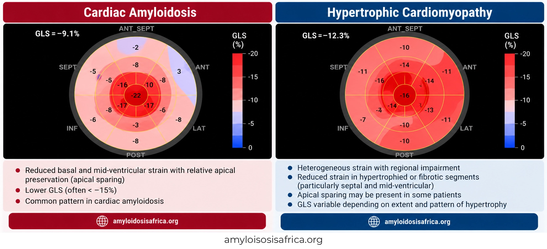
Figure 2. Global Longitudinal Strain in Cardiac Amyloidosis and Hypertrophic Cardiomyopathy. Cardiac amyloidosis frequently demonstrates reduced basal and mid-ventricular longitudinal strain with relative apical preservation (apical sparing). In contrast, hypertrophic cardiomyopathy (HCM) often produces heterogeneous regional strain abnormalities associated with myocardial hypertrophy and fibrosis. However, these patterns can overlap and should not be interpreted independently.
Cardiac MRI: How Late Gadolinium Enhancement Helps
Cardiac MRI provides detailed information about ventricular morphology, myocardial fibrosis, and tissue composition.
It becomes particularly valuable when echocardiographic findings overlap or when clinicians suspect an infiltrative cardiomyopathy rather than primary HCM.
The 2024 HCM guideline specifically recommends CMR for diagnostic clarification when echocardiography is inconclusive and supports its use when alternative infiltrative or storage diseases are suspected (2).
LGE in Hypertrophic Cardiomyopathy
HCM commonly produces patchy or focal late gadolinium enhancement, often in areas of maximal hypertrophy or at right ventricular insertion points.
This enhancement generally reflects myocardial replacement fibrosis.
Importantly, extensive LGE in HCM also contributes to risk assessment for adverse cardiac events, including ventricular arrhythmias.
LGE in Cardiac Amyloidosis
Cardiac amyloidosis more typically produces diffuse subendocardial or transmural LGE, often involving multiple ventricular segments.
Other characteristic findings include abnormal gadolinium kinetics and difficulty achieving normal myocardial nulling.
These findings reflect extensive extracellular amyloid infiltration and expansion of the interstitial space.
However, LGE patterns vary with disease stage and can overlap with other cardiomyopathies. Furthermore, CMR cannot reliably distinguish AL amyloidosis from ATTR amyloidosis by itself (4).
Native T1 Mapping and Extracellular Volume
CMR tissue mapping adds important diagnostic information beyond conventional LGE imaging.
Native T1
Cardiac amyloidosis commonly produces elevated native T1 values because of myocardial infiltration and extracellular expansion.
HCM may also increase native T1, particularly where fibrosis is present. However, the magnitude and distribution of abnormalities often differ.
Because native T1 values depend on scanner field strength, sequence, and local reference ranges, clinicians should avoid applying a single universal cutoff.
Extracellular Volume
Extracellular volume fraction, or ECV, estimates the proportion of myocardial tissue occupied by the extracellular compartment.
Cardiac amyloidosis frequently causes marked ECV expansion because amyloid fibrils accumulate extracellularly.
In HCM, ECV may increase because of interstitial fibrosis, but widespread marked expansion is generally more suggestive of infiltrative disease.
A meta-analysis by Pan and colleagues found that ECV mapping offered substantial diagnostic and prognostic value in cardiac amyloidosis (5).
| CMR Feature | Cardiac Amyloidosis | HCM |
|---|---|---|
| LGE distribution | Often diffuse subendocardial or transmural | Often patchy mid-wall or junctional |
| Native T1 | Frequently markedly elevated | May be elevated, especially with fibrosis |
| ECV | Frequently substantially increased | Variable increase, often less extensive |
| Myocardial nulling | May be abnormal | Usually lacks the characteristic amyloid nulling pattern |
| Apical aneurysm | Not characteristic | May occur, particularly in selected HCM phenotypes |
| Diagnostic limitation | Cannot establish AL versus ATTR subtype alone | CMR phenotype does not always establish a genetic cause |
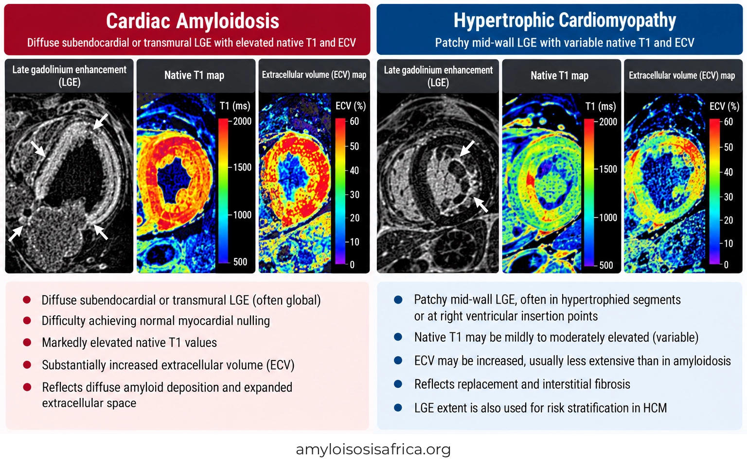
Figure 3. Cardiac MRI Differences Between Cardiac Amyloidosis and Hypertrophic Cardiomyopathy. Cardiac amyloidosis typically demonstrates diffuse subendocardial or transmural late gadolinium enhancement (LGE), elevated native T1 values, and markedly increased extracellular volume (ECV), reflecting myocardial amyloid infiltration. In contrast, hypertrophic cardiomyopathy (HCM) more commonly shows patchy mid-wall LGE in hypertrophied segments or right ventricular insertion regions, with variable increases in native T1 and ECV associated with myocardial fibrosis. Although these imaging characteristics help differentiate the conditions, findings may overlap and should be interpreted alongside clinical and other diagnostic investigations.
ECG Differences: Low Voltage vs Hypertrophy
The electrocardiogram may provide additional clues when clinicians evaluate unexplained ventricular wall thickening.
Cardiac amyloidosis can produce low QRS voltage despite increased wall thickness on echocardiography. This apparent discrepancy between electrical voltage and ventricular thickness is a classic diagnostic clue.
Other findings include atrial fibrillation, conduction abnormalities, pseudoinfarction patterns and atrioventricular block.
In contrast, HCM frequently produces high-voltage QRS complexes, repolarization abnormalities, deep T-wave inversion or pathological Q waves.
However, these patterns overlap. Notably, low QRS voltage is absent in many patients with ATTR-CM. Therefore, normal or increased voltage does not exclude cardiac amyloidosis.
Clinical Red Flags That Favor Cardiac Amyloidosis
Clinical history often helps distinguish cardiac amyloidosis from HCM before advanced imaging becomes available.
Features that should increase suspicion for cardiac amyloidosis include:
- older age at presentation, particularly with unexplained heart failure;
- bilateral carpal tunnel syndrome;
- lumbar spinal stenosis;
- atraumatic biceps tendon rupture;
- progressive peripheral neuropathy;
- autonomic dysfunction or orthostatic hypotension;
- unexplained proteinuria or nephrotic syndrome;
- macroglossia or periorbital purpura, particularly in AL amyloidosis;
- conduction disease or pacemaker implantation; and
- disproportionately elevated cardiac biomarkers.
By comparison, a family history of HCM, unexplained sudden cardiac death, or a known pathogenic sarcomeric variant may support HCM.
Nevertheless, hereditary ATTR can also produce familial cardiac disease. Therefore, family history alone cannot reliably distinguish the conditions.
Can Cardiac Amyloidosis Cause Asymmetric Septal Hypertrophy?
Yes. Although concentric wall thickening is often associated with cardiac amyloidosis, some patients develop asymmetric septal hypertrophy.
This is particularly relevant in ATTR cardiomyopathy, where myocardial morphology may resemble HCM.
Consequently, clinicians should investigate possible amyloidosis when asymmetric hypertrophy accompanies characteristic extracardiac red flags, abnormal strain, suspicious CMR findings or otherwise unexplained heart failure.
Can HCM Show Apical Sparing?
Yes. Relative apical sparing is not exclusive to cardiac amyloidosis.
Patients with HCM may demonstrate regional strain abnormalities that preserve apical function relative to more affected myocardial segments.
Therefore, clinicians should avoid diagnosing cardiac amyloidosis solely because the GLS bull’s-eye map shows a cherry-on-top pattern.
How Do Clinicians Confirm the Correct Diagnosis?
When echocardiography or CMR raises suspicion for cardiac amyloidosis, clinicians should follow a structured diagnostic pathway.
Step 1: Assess the Clinical Phenotype
Review age, symptoms, family history, hypertension, valve disease, neuropathy, orthopedic history, ECG findings and other potential causes of ventricular hypertrophy.
Step 2: Evaluate for AL Amyloidosis
When cardiac amyloidosis is suspected, obtain serum free light chains, serum immunofixation, and urine immunofixation.
This evaluation is essential because AL amyloidosis requires prompt hematological assessment and treatment.
Learn more about monoclonal protein testing in cardiac amyloidosis.
Step 3: Investigate ATTR Cardiomyopathy
In appropriate patients, bone-tracer scintigraphy using technetium-99m PYP, DPD, or HMDP can help establish ATTR cardiomyopathy.
When monoclonal protein testing is negative, and scintigraphy demonstrates grade 2 or 3 myocardial uptake with appropriate SPECT confirmation, clinicians may establish ATTR-CM noninvasively in the correct clinical setting.
However, when monoclonal protein testing is abnormal, scintigraphy alone cannot reliably distinguish ATTR from AL amyloidosis. Tissue biopsy with accurate amyloid typing may be necessary (6).
Read about bone scintigraphy and biopsy in cardiac amyloidosis.
Step 4: Evaluate for HCM
When HCM remains the leading diagnosis, clinicians should assess the full morphological phenotype, dynamic LVOT gradients, family history, and risk markers.
CMR may clarify ventricular hypertrophy patterns, identify apical abnormalities, and assess myocardial fibrosis.
Genetic counseling and testing can also support diagnosis and family screening when appropriate.
Step 5: Establish the Amyloid Subtype if ATTR Is Confirmed
Once clinicians confirm ATTR amyloidosis, TTR genetic testing distinguishes hereditary from wild-type disease.
This distinction has important implications for family counseling and genetic risk assessment.
Why Does the Correct Diagnosis Matter for Treatment?
Cardiac amyloidosis and HCM require different disease-specific management strategies.
Treatment of Cardiac Amyloidosis
ATTR-CM treatment may include disease-modifying therapies that stabilize transthyretin or reduce its production, depending on the clinical indication, regulatory approval, and patient characteristics.
In contrast, AL amyloidosis requires treatment directed at the underlying plasma-cell disorder.
Heart-failure and arrhythmia management must also account for the distinctive hemodynamic features of cardiac amyloidosis.
For more information, see cardiac amyloidosis treatment.
Treatment of HCM
HCM management may include beta-blockers, selected calcium-channel blockers, cardiac myosin inhibitors, or septal reduction therapy in appropriately selected patients with obstructive disease.
Clinicians also assess sudden cardiac death risk and determine whether an implantable cardioverter-defibrillator is indicated.
These interventions do not treat the underlying amyloid process. Consequently, diagnostic misclassification can delay appropriate disease-modifying treatment.
Cardiac Amyloidosis vs HCM: Summary Comparison
| Diagnostic Domain | Cardiac Amyloidosis | HCM |
|---|---|---|
| Underlying mechanism | Extracellular amyloid infiltration | Primary cardiomyocyte hypertrophy, often sarcomeric |
| Typical morphology | Often diffuse wall thickening | Often asymmetric septal hypertrophy |
| LVOTO | Less typical | Characteristic of obstructive HCM |
| GLS pattern | Relative apical sparing often present | Regional impairment often associated with hypertrophy |
| CMR LGE | Often diffuse subendocardial or transmural | Often patchy mid-wall or junctional |
| ECV | Frequently markedly elevated | Variable elevation |
| ECG | Low voltage, pseudoinfarction or conduction disease may occur | LVH voltage, repolarization abnormalities or pathological Q waves |
| Extracardiac clues | Carpal tunnel, neuropathy, autonomic or systemic manifestations | Family history of HCM or sudden cardiac death may be present |
| Confirmatory evaluation | Monoclonal testing, scintigraphy, biopsy and typing when indicated | Clinical imaging phenotype, family evaluation and selected genetic testing |
Why This Distinction Matters in Africa
In many African healthcare settings, echocardiography is more accessible than cardiac MRI, nuclear scintigraphy, or specialized amyloid pathology.
Consequently, clinicians may initially classify patients with unexplained ventricular thickening as having hypertensive heart disease or HCM without fully evaluating infiltrative causes.
However, the true frequency of missed cardiac amyloidosis among patients labeled with HCM across African populations remains insufficiently characterized.
Improving diagnostic recognition requires:
- greater awareness of amyloidosis red flags;
- systematic evaluation of unexplained ventricular hypertrophy;
- appropriate use of echocardiographic strain imaging;
- access to monoclonal protein testing;
- referral pathways for advanced imaging and amyloid typing;
- improved access to genetic evaluation; and
- stronger African multicenter research and registry data.
Learn more about cardiac amyloidosis in Africa.
Key Takeaways
- Cardiac amyloidosis and HCM can both cause ventricular hypertrophy, diastolic dysfunction, and heart failure.
- Cardiac amyloidosis results from extracellular amyloid deposition, while HCM generally involves primary myocardial hypertrophy.
- HCM often produces asymmetric septal hypertrophy, but cardiac amyloidosis may also show asymmetric patterns.
- Relative apical sparing on GLS is an important amyloidosis clue but is not specific or diagnostic.
- CMR often demonstrates diffuse subendocardial or transmural LGE in amyloidosis, compared with more focal or patchy fibrosis in HCM.
- Native T1 and ECV mapping provide additional information about myocardial tissue composition.
- Clinical red flags and family history can help direct investigations.
- Suspected cardiac amyloidosis requires appropriate testing to distinguish AL from ATTR.
- Accurate diagnosis is essential because treatment strategies differ substantially.
Frequently Asked Questions
Can cardiac amyloidosis be mistaken for HCM?
Yes. Both conditions can produce increased ventricular wall thickness, impaired diastolic function and abnormal myocardial strain. Additional clinical and imaging investigations are often necessary.
Does asymmetric septal hypertrophy always indicate HCM?
No. Although asymmetric septal hypertrophy is common in HCM, it can also occur in cardiac amyloidosis, particularly ATTR-CM.
Is apical sparing specific for cardiac amyloidosis?
No. Relative apical sparing increases suspicion for cardiac amyloidosis but can also occur in other cardiac conditions.
Which CMR finding favors cardiac amyloidosis?
Diffuse subendocardial or transmural LGE, elevated native T1, markedly expanded ECV and abnormal myocardial nulling support suspicion for cardiac amyloidosis.
Can HCM produce elevated native T1?
Yes. Myocardial fibrosis in HCM can increase native T1 and ECV, although the distribution and degree of abnormalities may differ from amyloidosis.
Can echocardiography alone distinguish amyloidosis from HCM?
Not reliably in every patient. Echocardiography provides valuable clues, but some patients require CMR, laboratory testing, scintigraphy, genetic evaluation or biopsy.
Does cardiac MRI confirm ATTR amyloidosis?
No. CMR can strongly suggest cardiac amyloid infiltration, but it does not reliably establish whether the amyloid is ATTR or AL.
Can HCM and cardiac amyloidosis coexist?
Yes, although coexistence is uncommon. Clinicians should consider more than one diagnosis when the clinical and imaging findings cannot be explained by a single condition.
Why is distinguishing these conditions important?
The diseases require different treatments, risk assessments, and follow-up strategies. Accurate classification can prevent inappropriate treatment and reduce diagnostic delays.
References
- Garcia-Pavia P, Rapezzi C, Adler Y, et al. Diagnosis and treatment of cardiac amyloidosis: a position statement of the ESC Working Group on Myocardial and Pericardial Diseases. Eur Heart J. 2021;42(16):1554–1568. doi:10.1093/eurheartj/ehab072.
- Ommen SR, Ho CY, Asif IM, et al. 2024 AHA/ACC/AMSSM/HRS/PACES/SCMR Guideline for the Management of Hypertrophic Cardiomyopathy. J Am Coll Cardiol. 2024. doi:10.1016/j.jacc.2024.02.014.
- Phelan D, Collier P, Thavendiranathan P, et al. Relative apical sparing of longitudinal strain using two-dimensional speckle-tracking echocardiography is both sensitive and specific for the diagnosis of cardiac amyloidosis. Heart. 2012;98(19):1442–1448. doi:10.1136/heartjnl-2012-302353.
- How to Image Cardiac Amyloidosis: A Practical Approach. JACC Cardiovasc Imaging. View publication.
- Pan JA, Kerwin MJ, Salerno M. Native T1 Mapping, Extracellular Volume Mapping, and Late Gadolinium Enhancement in Cardiac Amyloidosis: A Meta-Analysis. JACC Cardiovasc Imaging. 2020. doi:10.1016/j.jcmg.2020.03.010.
- Gillmore JD, Maurer MS, Falk RH, et al. Nonbiopsy diagnosis of cardiac transthyretin amyloidosis. Circulation. 2016;133(24):2404–2412. doi:10.1161/CIRCULATIONAHA.116.021612.
- Kittleson MM, Ruberg FL, Ambardekar AV, et al. 2023 ACC Expert Consensus Decision Pathway on Comprehensive Multidisciplinary Care for the Patient With Cardiac Amyloidosis. J Am Coll Cardiol. 2023;81(11):1076–1126. doi:10.1016/j.jacc.2022.11.022.
This article provides educational information for healthcare professionals and the public. It does not replace individualized medical assessment, diagnostic investigations or specialist recommendations. Cardiac amyloidosis and hypertrophic cardiomyopathy may have overlapping clinical and imaging features, and definitive diagnosis requires appropriate clinical evaluation. Diagnostic and treatment decisions should follow current professional guidelines and local regulatory requirements.



Leave a Reply