



Cardiac Amyloidosis vs Fabry Disease: How to Tell Them Apart
Cardiac amyloidosis vs Fabry disease is an important diagnostic comparison because both conditions can cause increased left ventricular wall thickness, diastolic dysfunction, abnormal myocardial strain, arrhythmias and heart failure.
Despite these similarities, the two diseases have fundamentally different causes, diagnostic tests and treatment approaches.
Cardiac amyloidosis results from extracellular deposition of misfolded proteins within the myocardium. In contrast, Fabry disease is an inherited lysosomal storage disorder caused by deficient alpha-galactosidase A activity, leading to intracellular accumulation of glycosphingolipids.
Both conditions may initially be mistaken for hypertrophic cardiomyopathy or hypertensive heart disease, especially when ventricular hypertrophy is the dominant imaging finding.
However, cardiac magnetic resonance imaging (CMR), characteristic extracardiac manifestations, biochemical testing, and genetic investigations can help distinguish them.
This article explains the key differences between cardiac amyloidosis and Fabry disease, focusing on clinical presentation, echocardiography, global longitudinal strain (GLS), CMR, laboratory testing and treatment.
What Is Cardiac Amyloidosis?
Cardiac amyloidosis is an infiltrative cardiomyopathy caused by deposition of abnormal amyloid fibrils in the myocardial extracellular space.
The two principal types are:
- Transthyretin amyloidosis (ATTR): caused by deposition of misfolded transthyretin, including hereditary ATTR (ATTRv) and wild-type ATTR (ATTRwt).
- Immunoglobulin light-chain amyloidosis (AL): associated with a plasma-cell disorder producing amyloidogenic immunoglobulin light chains.
As amyloid accumulates, ventricular walls may appear thicker, myocardial relaxation becomes impaired, and progressive cardiac dysfunction may develop.
Patients may present with heart failure, atrial fibrillation, conduction abnormalities, exertional intolerance, or characteristic extracardiac manifestations.
Read more about hereditary ATTR amyloidosis, wild-type ATTR amyloidosis and AL amyloidosis.
What Is Fabry Disease?
Fabry disease is an X-linked lysosomal storage disorder caused by pathogenic variants in the GLA gene.
These variants may reduce or abolish alpha-galactosidase A activity, resulting in accumulation of globotriaosylceramide (Gb3) and related glycosphingolipids in multiple tissues.
Fabry disease can affect the:
- heart;
- kidneys;
- peripheral and autonomic nervous systems;
- skin;
- eyes; and
- cerebrovascular system.
Cardiac involvement commonly includes left ventricular hypertrophy, myocardial fibrosis, conduction abnormalities and arrhythmias.
Although Fabry disease is X-linked, women with pathogenic GLA variants may develop substantial cardiac and extracardiac disease. The severity and age of onset are variable.
Some individuals develop classic multisystem disease early in life, while others have later-onset phenotypes with predominantly cardiac manifestations.
Why Can Fabry Disease Mimic Cardiac Amyloidosis?
Both conditions can cause concentric left ventricular wall thickening, impaired diastolic function and reduced myocardial longitudinal strain.
Moreover, both may be associated with conduction disease, arrhythmias, and progressive heart failure.
However, the mechanisms of myocardial thickening differ.
Cardiac Amyloidosis: Extracellular Infiltration
Amyloid fibrils accumulate between myocardial cells, expanding the extracellular compartment.
This can produce substantial extracellular volume expansion, abnormal gadolinium kinetics, and characteristic CMR enhancement patterns.
Fabry Disease: Intracellular Storage
In Fabry disease, glycosphingolipids accumulate predominantly within lysosomes of affected cells.
This intracellular storage contributes to myocardial hypertrophy and may produce reduced native T1 values on CMR, particularly before extensive fibrosis develops.
Over time, inflammation and replacement fibrosis can complicate the imaging appearance.
Echocardiography: Similar Hypertrophy, Different Disease
Echocardiography is frequently the first imaging investigation to identify cardiac involvement in either condition.
Cardiac Amyloidosis
Typical findings may include:
- increased left ventricular wall thickness;
- biatrial enlargement;
- right ventricular wall thickening;
- thickening of atrioventricular valves or the interatrial septum;
- diastolic dysfunction; and
- reduced longitudinal strain.
However, these findings vary with disease stage and amyloid subtype.
Fabry Disease
Fabry cardiomyopathy commonly demonstrates:
- concentric left ventricular hypertrophy;
- preserved ejection fraction in earlier stages;
- diastolic dysfunction;
- papillary muscle hypertrophy in some patients;
- reduced myocardial strain; and
- left atrial enlargement with disease progression.
Asymmetric hypertrophy may also occur in Fabry disease.
Consequently, echocardiographic morphology alone cannot reliably distinguish Fabry disease from cardiac amyloidosis.
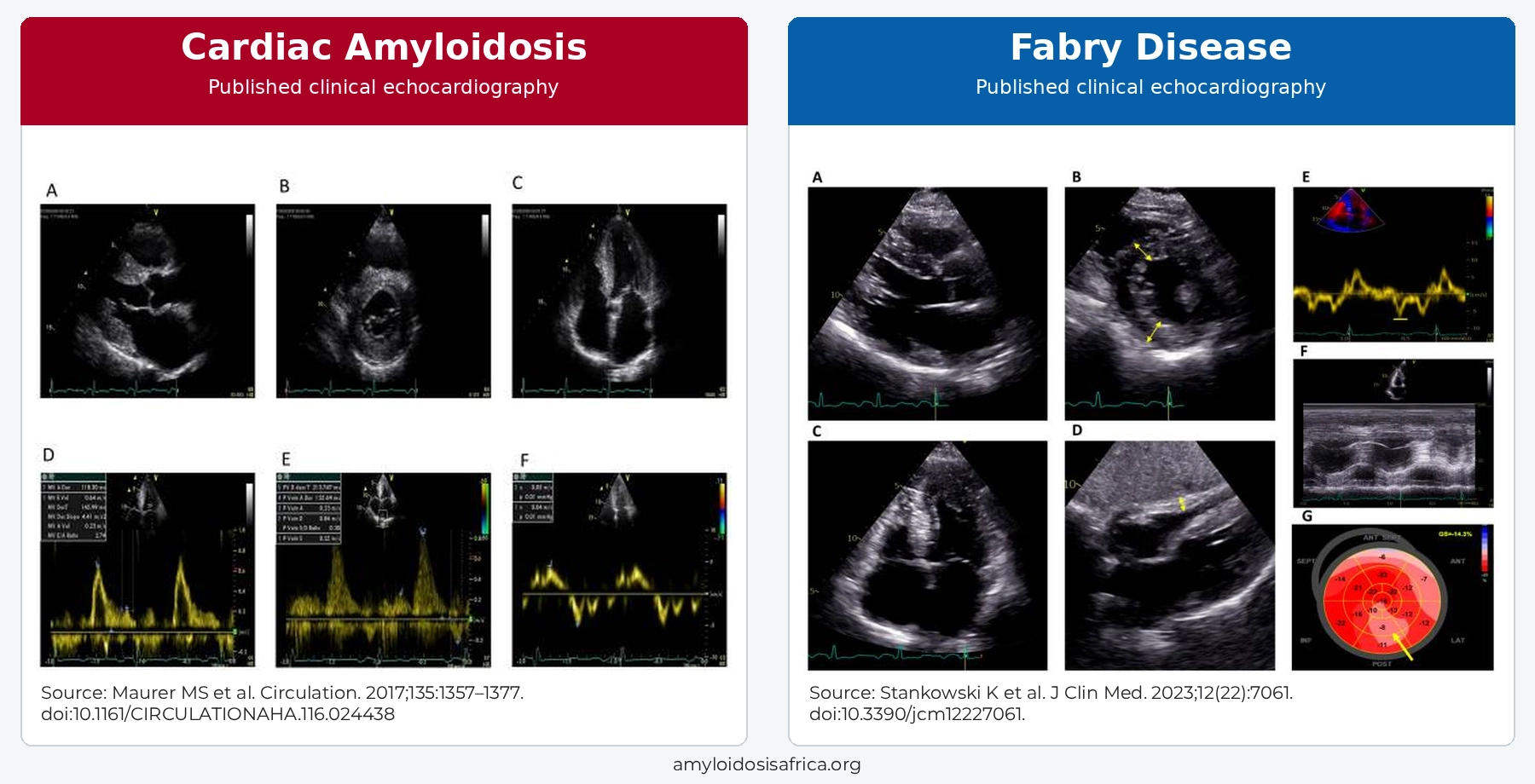
Figure 1. Echocardiographic Differences Between Cardiac Amyloidosis and Fabry Disease. Published clinical echocardiographic images illustrating cardiac involvement in both conditions. Cardiac amyloidosis (left) may demonstrate increased ventricular wall thickness, biatrial enlargement, and abnormalities of myocardial longitudinal strain. Fabry disease (right) commonly presents with left ventricular hypertrophy, sometimes accompanied by papillary muscle enlargement and regional strain impairment. Although these findings provide diagnostic clues, substantial overlap exists, and echocardiography alone cannot reliably distinguish the two conditions.
Global Longitudinal Strain: Does Apical Sparing Help?
Global longitudinal strain measures myocardial deformation and may detect impaired systolic function before the left ventricular ejection fraction declines.
GLS in Cardiac Amyloidosis
Cardiac amyloidosis frequently produces impaired basal and mid-ventricular longitudinal strain with relative preservation of apical strain.
This pattern is commonly described as relative apical sparing.
However, it is not specific to amyloidosis and may be absent in some patients.
GLS in Fabry Disease
Fabry cardiomyopathy may produce reduced longitudinal strain, including regional impairment associated with hypertrophy and fibrosis.
Reduced strain in the basal inferolateral region can be particularly relevant because this region is frequently affected by replacement fibrosis in Fabry disease.
Nevertheless, GLS patterns overlap, and neither regional strain impairment nor apical sparing establishes the diagnosis independently.
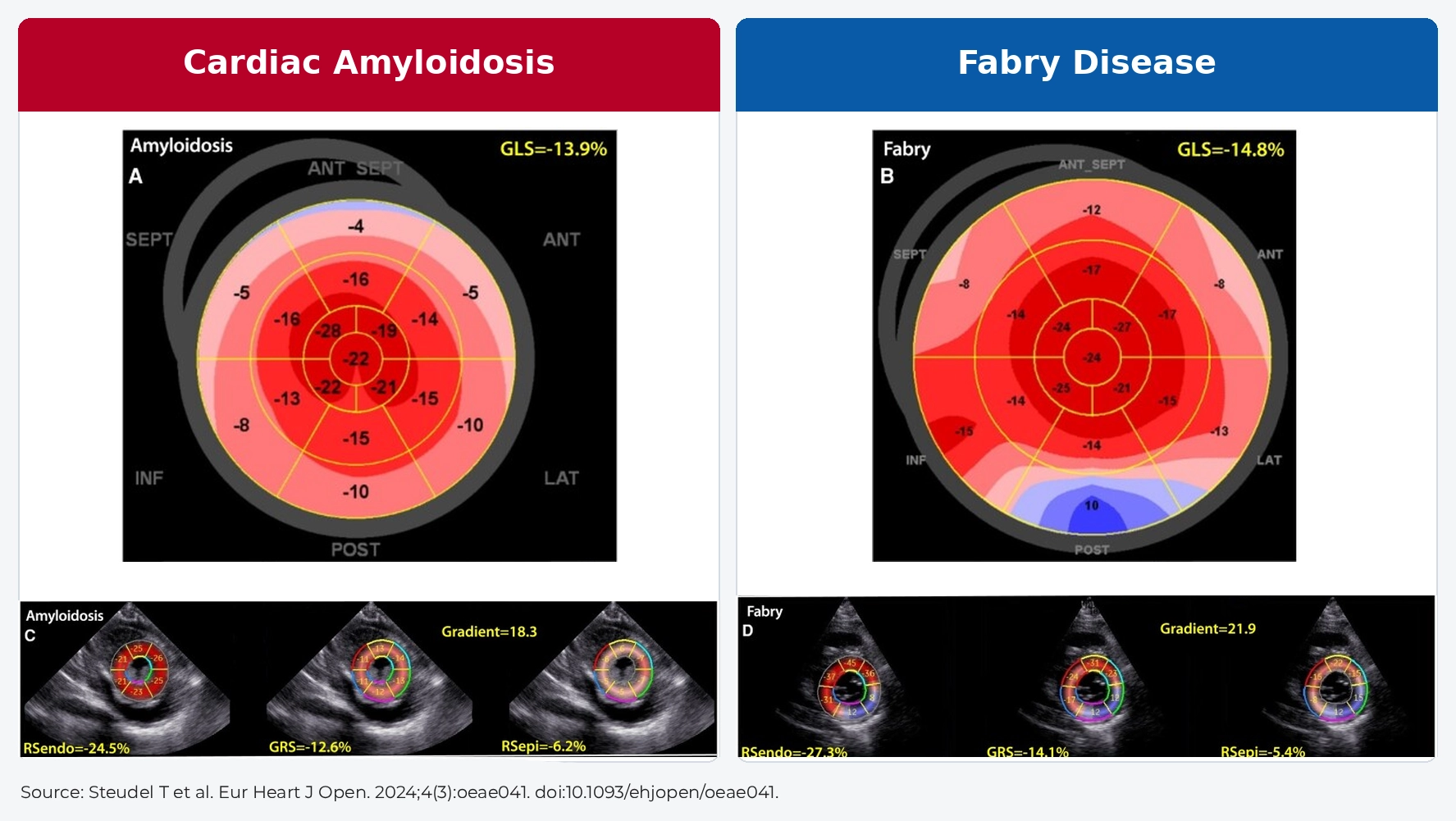
Figure 2. Global Longitudinal Strain in Cardiac Amyloidosis and Fabry Disease. Published speckle-tracking echocardiographic images comparing myocardial deformation patterns in cardiac amyloidosis and Fabry disease. Both conditions demonstrate impaired global longitudinal strain (GLS), with reported values of −13.9% and −14.8%, respectively. Cardiac amyloidosis frequently exhibits relative apical sparing, whereas Fabry cardiomyopathy may show regional strain impairment, particularly in the basal inferolateral segments. However, these patterns can overlap and are not independently diagnostic.
Cardiac MRI: One of the Most Useful Differentiating Tools
Cardiac MRI is particularly valuable because it provides information about myocardial morphology, fibrosis, and tissue composition.
The combination of late gadolinium enhancement (LGE), native T1 mapping, and extracellular volume (ECV) estimation may help distinguish Fabry disease from cardiac amyloidosis.
Native T1 Mapping: A Key Difference
One of the most characteristic distinctions is the direction of native T1 change.
Cardiac amyloidosis: Native T1 is commonly elevated because of extracellular amyloid deposition and associated myocardial changes.
Fabry disease: Native T1 is often reduced in affected myocardium because of intracellular glycosphingolipid accumulation.
However, reduced T1 is not universal in Fabry disease. Native T1 may become normal or elevated in regions with substantial inflammation or fibrosis, especially in advanced disease.
Similarly, elevated native T1 is not specific to amyloidosis and may occur in several other myocardial diseases.
Late Gadolinium Enhancement
Cardiac amyloidosis frequently demonstrates diffuse subendocardial or transmural LGE, sometimes accompanied by abnormal myocardial nulling.
In Fabry disease, LGE commonly develops in the basal inferolateral wall, often with a mid-wall distribution.
However, Fabry disease can demonstrate other enhancement patterns, and amyloidosis may present with variable LGE distributions.
Therefore, LGE distribution should be interpreted as part of a comprehensive tissue-characterization assessment.
Extracellular Volume
ECV is often markedly elevated in cardiac amyloidosis because amyloid deposits expand the myocardial extracellular space.
In Fabry disease, ECV may remain normal in myocardium without fibrosis because the primary storage process is intracellular.
However, ECV can increase in fibrotic regions and in advanced Fabry cardiomyopathy.
Consequently, ECV findings must be interpreted alongside native T1, LGE, and the overall clinical picture.
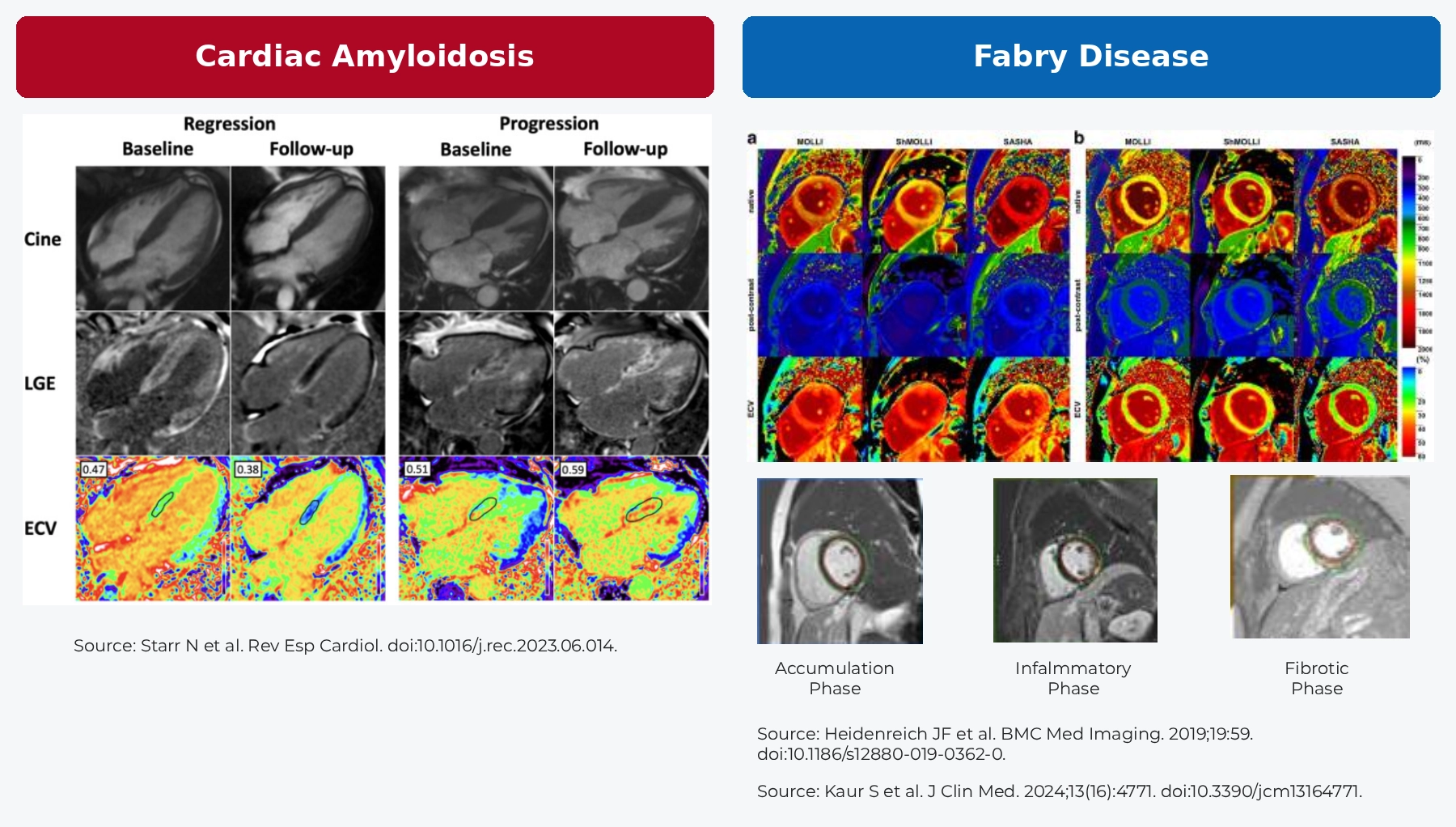
Figure 3. Cardiac MRI Differences Between Cardiac Amyloidosis and Fabry Disease. Published cardiac magnetic resonance (CMR) images illustrating differences in myocardial tissue characteristics. Cardiac amyloidosis commonly demonstrates diffuse subendocardial or transmural late gadolinium enhancement (LGE), elevated native T1, and substantial extracellular volume (ECV) expansion. In contrast, Fabry cardiomyopathy often exhibits reduced native T1 due to intracellular glycosphingolipid accumulation, with LGE frequently involving the basal inferolateral wall. Advanced fibrosis may alter these patterns, so interpret findings alongside clinical, biochemical, and genetic investigations.
Clinical Red Flags: Amyloidosis vs Fabry Disease
Extracardiac manifestations are particularly helpful when evaluating patients with unexplained left ventricular hypertrophy.
Findings Suggesting Cardiac Amyloidosis
- bilateral carpal tunnel syndrome;
- lumbar spinal stenosis;
- atraumatic biceps tendon rupture;
- peripheral or autonomic neuropathy;
- orthostatic hypotension;
- unexplained heart failure with increased ventricular wall thickness; and
- proteinuria or nephrotic syndrome, particularly in AL amyloidosis.
Some manifestations differ according to amyloid subtype. For example, macroglossia and periorbital purpura suggest AL amyloidosis more than ATTR-CM.
Findings Suggesting Fabry Disease
- neuropathic pain or burning sensations in the hands and feet (acroparesthesias);
- reduced sweating or heat intolerance;
- angiokeratomas;
- cornea verticillata;
- proteinuria or progressive kidney dysfunction;
- early-onset stroke or transient ischemic attack;
- gastrointestinal symptoms, including abdominal pain or diarrhea; and
- a family history compatible with X-linked inheritance.
Importantly, later-onset cardiac Fabry disease may occur without many of the classic childhood manifestations.
Similarly, the absence of extracardiac red flags does not exclude cardiac amyloidosis.
Comparison Table: Cardiac Amyloidosis vs Fabry Disease
| Feature | Cardiac Amyloidosis | Fabry Disease |
|---|---|---|
| Underlying mechanism | Extracellular amyloid deposition | Intracellular glycosphingolipid storage |
| Principal cause | ATTR or AL amyloid proteins | Pathogenic variants in the GLA gene |
| Inheritance | ATTRv is autosomal dominant; ATTRwt and AL are not inherited in the same manner | X-linked |
| Typical cardiac presentation | Infiltrative cardiomyopathy and heart failure | Hypertrophic phenotype, fibrosis and arrhythmias |
| LV wall thickening | Common | Common |
| GLS | Often reduced with relative apical sparing | Regional impairment, sometimes basal inferolateral |
| Native T1 | Usually elevated | Often reduced before extensive fibrosis |
| ECV | Frequently substantially elevated | Often normal outside fibrotic regions |
| LGE | Often diffuse subendocardial or transmural | Often basal inferolateral mid-wall |
| Characteristic extracardiac clues | Carpal tunnel syndrome, neuropathy, autonomic dysfunction and subtype-dependent manifestations | Acroparesthesias, angiokeratomas, cornea verticillata, kidney disease and early stroke |
| Diagnostic confirmation | Subtype-specific monoclonal testing, bone scintigraphy or amyloid biopsy | Alpha-galactosidase A activity and GLA genetic testing |
| Disease-specific treatment | ATTR-directed therapy or AL-directed plasma-cell treatment | Enzyme replacement therapy or eligible mutation-specific pharmacological chaperone therapy |
How Is Cardiac Amyloidosis Diagnosed?
When cardiac amyloidosis is suspected, determining the amyloid subtype is essential.
Step 1: Screen for Monoclonal Proteins
The initial laboratory assessment should include:
- serum free light chains with kappa/lambda ratio;
- serum immunofixation; and
- urine immunofixation.
These investigations help evaluate the possibility of AL amyloidosis.
Serum or urine protein electrophoresis alone is insufficient to exclude AL amyloidosis.
Learn more about monoclonal protein testing in cardiac amyloidosis.
Step 2: Assess for ATTR-CM
When the monoclonal protein screen is negative, technetium-99m PYP, DPD or HMDP bone scintigraphy with SPECT may support a nonbiopsy diagnosis of ATTR-CM.
In the appropriate clinical setting, grade 2 or 3 myocardial tracer uptake together with a negative monoclonal protein screen can establish ATTR-CM noninvasively.
However, positive monoclonal testing, equivocal imaging or discordant findings may require tissue biopsy with accurate amyloid typing.
Learn more about bone scintigraphy and biopsy in cardiac amyloidosis.
Step 3: Perform TTR Genetic Testing
Once ATTR amyloidosis is established, TTR genetic testing helps distinguish hereditary from wild-type disease and guides family counseling.
How Is Fabry Disease Diagnosed?
Fabry disease requires disease-specific biochemical and molecular investigations.
Alpha-Galactosidase A Enzyme Activity
In males with classic Fabry disease, alpha-galactosidase A activity is often markedly reduced.
However, residual enzyme activity may occur in later-onset phenotypes.
Importantly, enzyme activity can be within the normal range in females with Fabry disease. Therefore, normal enzyme activity does not exclude Fabry disease in women.
GLA Genetic Testing
Molecular testing of the GLA gene is essential for confirming a genetic diagnosis, particularly in females and individuals with atypical presentations.
However, identification of a variant of uncertain significance does not establish Fabry disease on its own.
Variant interpretation should consider clinical findings, enzyme activity, biomarkers, family segregation and established classification criteria.
Lyso-Gb3 Testing
Plasma globotriaosylsphingosine (lyso-Gb3) can support diagnosis and disease assessment.
However, biomarker concentrations vary by phenotype and sex, and a normal or minimally elevated result does not reliably exclude Fabry disease.
Specialist interpretation is therefore important.
Diagnostic Approach to Unexplained Left Ventricular Hypertrophy
When increased ventricular wall thickness cannot be adequately explained by hypertension, aortic stenosis or another established condition, clinicians should consider infiltrative and storage disorders.
- Review the clinical phenotype: Assess age, family history, cardiac symptoms, kidney disease, neuropathy, skin findings and other extracardiac manifestations.
- Evaluate ECG and echocardiography: Examine ventricular geometry, diastolic function, conduction abnormalities and longitudinal strain.
- Perform cardiac MRI when appropriate: Assess native T1, ECV and LGE patterns for clues to myocardial infiltration or storage disease.
- Investigate suspected amyloidosis: Obtain monoclonal protein testing and pursue subtype-appropriate scintigraphy or biopsy.
- Investigate suspected Fabry disease: Perform alpha-galactosidase A activity testing where appropriate, GLA genetic testing and supportive biomarker evaluation.
- Integrate results: Confirm the diagnosis through appropriate specialist assessment before initiating disease-specific therapy.
Importantly, imaging findings alone should not be used to label a patient as having either disease.
Why the Correct Diagnosis Changes Treatment
Treatment of Cardiac Amyloidosis
Treatment depends on the underlying amyloid protein.
ATTR-CM may be treated with approved transthyretin stabilizers or TTR-directed therapies, depending on the patient’s indication and local availability.
AL amyloidosis requires prompt specialist evaluation and treatment directed at the underlying plasma-cell disorder.
Supportive management of heart failure, arrhythmias and conduction disease is also important.
Read more about cardiac amyloidosis treatment.
Treatment of Fabry Disease
Fabry-specific treatment may include:
- Enzyme replacement therapy: Provides recombinant alpha-galactosidase A to reduce glycosphingolipid accumulation.
- Pharmacological chaperone therapy: Migalastat may be appropriate for patients with eligible amenable GLA variants, subject to regulatory indications and clinical suitability.
Supportive care may also address kidney disease, arrhythmias, heart failure, neuropathic pain and cerebrovascular risk.
Established myocardial fibrosis may limit the extent of cardiac recovery despite disease-specific treatment.
Therefore, early recognition and multidisciplinary follow-up are important.
Why This Distinction Matters in Africa
Both cardiac amyloidosis and Fabry disease are likely to be underrecognized in many African healthcare settings, although robust population-level estimates remain limited.
Patients with unexplained ventricular hypertrophy may initially be classified as having hypertensive heart disease or hypertrophic cardiomyopathy without investigation for infiltrative or storage disorders.
Diagnostic challenges may include limited access to:
- advanced echocardiographic strain analysis;
- cardiac MRI with T1 and ECV mapping;
- alpha-galactosidase A enzyme assays;
- GLA and TTR genetic testing;
- bone scintigraphy;
- specialist pathology and amyloid typing; and
- disease-specific treatment.
Increasing clinician awareness and establishing appropriate referral pathways could support earlier recognition of these conditions.
In addition, multicenter African research may help clarify disease presentation, diagnostic access and treatment gaps.
Learn more about cardiac amyloidosis in Africa.
Key Takeaways
- Cardiac amyloidosis and Fabry disease can both cause increased left ventricular wall thickness, diastolic dysfunction and abnormal myocardial strain.
- Cardiac amyloidosis involves extracellular amyloid deposition, whereas Fabry disease causes intracellular glycosphingolipid accumulation.
- Cardiac amyloidosis typically increases native T1 and ECV, while Fabry disease often reduces native T1 before extensive fibrosis develops.
- Diffuse subendocardial or transmural LGE favors cardiac amyloidosis, whereas basal inferolateral mid-wall LGE is characteristic of Fabry cardiomyopathy.
- Neither echocardiography nor cardiac MRI can independently confirm the diagnosis.
- Fabry disease requires appropriate biochemical and GLA genetic investigations.
- Cardiac amyloidosis requires accurate identification of AL or ATTR disease.
- Early diagnosis matters because treatment strategies differ substantially.
Frequently Asked Questions
Can Fabry disease be mistaken for cardiac amyloidosis?
Yes. Both conditions may cause left ventricular wall thickening, diastolic dysfunction, arrhythmias and abnormal myocardial strain. Cardiac MRI tissue mapping and disease-specific laboratory investigations can help distinguish them.
What is the main difference between Fabry disease and cardiac amyloidosis?
Fabry disease is an inherited lysosomal storage disorder involving intracellular glycosphingolipid accumulation. Cardiac amyloidosis results from extracellular deposition of misfolded amyloid proteins.
Does Fabry disease cause low native T1 on cardiac MRI?
Fabry disease frequently causes reduced native myocardial T1, particularly before extensive fibrosis develops. However, advanced myocardial fibrosis and inflammation may alter this pattern.
Is native T1 elevated in cardiac amyloidosis?
Native T1 is commonly elevated in cardiac amyloidosis because of extracellular amyloid deposition and associated myocardial changes. However, elevated native T1 is not specific to amyloidosis.
Can global longitudinal strain distinguish Fabry disease from amyloidosis?
GLS can provide supportive diagnostic clues, but strain patterns overlap. Relative apical sparing is not exclusive to cardiac amyloidosis, and disease-specific testing remains necessary.
Can women develop Fabry cardiomyopathy?
Yes. Women with pathogenic GLA variants can develop significant cardiac disease. Enzyme activity may be normal, making genetic testing particularly important.
Can Fabry disease cause kidney disease and neuropathy?
Yes. Fabry disease can affect the kidneys and peripheral nervous system, sometimes before cardiac manifestations become apparent.
Can cardiac amyloidosis and Fabry disease be treated?
Yes. Both have disease-specific treatment options, but the therapies are fundamentally different. Accurate diagnosis is essential before treatment selection.
References
- Garcia-Pavia P, Rapezzi C, Adler Y, et al. Diagnosis and treatment of cardiac amyloidosis: a position statement of the ESC Working Group on Myocardial and Pericardial Diseases. Eur Heart J. 2021;42(16):1554–1568. doi:10.1093/eurheartj/ehab072.
- Gillmore JD, Maurer MS, Falk RH, et al. Nonbiopsy Diagnosis of Cardiac Transthyretin Amyloidosis. Circulation. 2016;133(24):2404–2412. doi:10.1161/CIRCULATIONAHA.116.021612.
- Kittleson MM, Ruberg FL, Ambardekar AV, et al. 2023 ACC Expert Consensus Decision Pathway on Comprehensive Multidisciplinary Care for the Patient With Cardiac Amyloidosis. J Am Coll Cardiol. 2023;81(11):1076–1126. doi:10.1016/j.jacc.2022.11.022.
- Germain DP. Fabry disease. Orphanet J Rare Dis. 2010;5:30. doi:10.1186/1750-1172-5-30.
- Sado DM, White SK, Piechnik SK, et al. Identification and assessment of Anderson-Fabry disease by cardiovascular magnetic resonance noncontrast myocardial T1 mapping. Circ Cardiovasc Imaging. 2013;6(3):392–398. doi:10.1161/CIRCIMAGING.112.000070.
- Linhart A, Germain DP, Olivotto I, et al. An expert consensus document on the management of cardiovascular manifestations of Fabry disease. Eur J Heart Fail. 2020;22(7):1076–1096. doi:10.1002/ejhf.1960.
- Pieroni M, Moon JC, Arbustini E, et al. Cardiac involvement in Fabry disease: JACC Review Topic of the Week. J Am Coll Cardiol. 2021;77(7):922–936. doi:10.1016/j.jacc.2020.12.024.
This article is intended for educational purposes and does not replace individualized medical advice, specialist evaluation or current clinical guidelines. Cardiac amyloidosis and Fabry disease may have overlapping clinical and imaging findings. Diagnostic confirmation and treatment decisions should be made by qualified healthcare professionals using appropriate biochemical, genetic, imaging and pathological investigations.

Leave a Reply