



Cardiac Amyloidosis vs Aortic Stenosis: How to Tell Them Apart
Cardiac amyloidosis vs aortic stenosis is an important clinical comparison because both conditions can cause left ventricular wall thickening, diastolic dysfunction, reduced myocardial strain and heart failure.
However, the diagnostic challenge extends beyond distinguishing one disease from another. Aortic stenosis and cardiac amyloidosis can occur together, particularly in older adults with transthyretin amyloid cardiomyopathy (ATTR-CM).
In such patients, pressure overload from a narrowed aortic valve and myocardial infiltration from amyloid deposits may contribute simultaneously to ventricular dysfunction.
Recognizing this coexistence is essential because treating the valve obstruction does not eliminate the underlying amyloid cardiomyopathy.
This article explains how clinical findings, echocardiography, global longitudinal strain (GLS), cardiac magnetic resonance imaging (CMR), bone scintigraphy and other investigations help clinicians distinguish these conditions and identify patients who may have both diseases.
What Is Aortic Stenosis?
Aortic stenosis (AS) is a condition in which narrowing of the aortic valve obstructs blood flow from the left ventricle into the aorta.
In older adults, calcific degeneration is a common cause. Other causes include congenital bicuspid aortic valve disease and rheumatic valvular disease.
As the valve becomes progressively narrowed, the left ventricle must generate higher pressure to maintain forward blood flow.
This chronic pressure overload may produce:
- concentric left ventricular hypertrophy;
- increased myocardial mass;
- diastolic dysfunction;
- myocardial fibrosis;
- reduced longitudinal myocardial strain;
- left atrial enlargement; and
- heart failure.
Patients may experience exertional breathlessness, chest discomfort, reduced exercise tolerance, presyncope or syncope.
However, symptoms may be subtle in older adults or attributed to other cardiovascular conditions.
What Is Cardiac Amyloidosis?
Cardiac amyloidosis occurs when misfolded proteins accumulate as amyloid fibrils within the myocardial extracellular space.
These deposits increase apparent ventricular wall thickness and impair myocardial relaxation and contraction.
The two principal types are:
- Transthyretin amyloidosis (ATTR): caused by deposition of misfolded transthyretin, including wild-type and hereditary forms.
- Immunoglobulin light-chain amyloidosis (AL): caused by abnormal immunoglobulin light chains associated with a plasma-cell disorder.
Wild-type ATTR-CM is particularly relevant in older patients undergoing evaluation for degenerative aortic stenosis.
Learn more about wild-type ATTR amyloidosis, hereditary ATTR amyloidosis and AL amyloidosis.
Why Do Aortic Stenosis and Cardiac Amyloidosis Look Similar?
Both diseases can cause increased ventricular wall thickness and impaired diastolic function. Moreover, both may produce heart failure despite a relatively preserved left ventricular ejection fraction.
However, their underlying mechanisms differ.
- Aortic stenosis: increased ventricular afterload produces cardiomyocyte hypertrophy and myocardial fibrosis.
- Cardiac amyloidosis: extracellular amyloid deposition causes infiltrative myocardial disease and increased extracellular volume.
In patients with both diseases, these mechanisms may act together, resulting in more complex ventricular remodeling and impaired forward flow.
How Common Is Cardiac Amyloidosis in Patients With Aortic Stenosis?
Published studies suggest that ATTR-CM is not uncommon among older adults referred for transcatheter aortic valve replacement (TAVR).
In a prospective multicenter study involving 407 patients referred for TAVR, Nitsche and colleagues identified positive bone scintigraphy in 11.8% of participants. Grade 2 or 3 uptake, supporting a diagnosis of ATTR-CM in the appropriate diagnostic setting, was observed in 7.9% (2).
These findings demonstrate that coexisting amyloid disease deserves attention during evaluation of older patients with severe aortic stenosis.
However, prevalence estimates vary across studies because of differences in age, patient selection, diagnostic methods, and definitions.
Therefore, these figures should not be generalized to all patients with aortic stenosis or to the wider African population.
Echocardiography: Distinguishing Valve Obstruction From Myocardial Infiltration
Transthoracic echocardiography is the primary imaging modality for evaluating aortic stenosis.
It provides information about aortic valve morphology, transvalvular velocity, pressure gradient, valve area, ventricular geometry and cardiac function.
Typical Findings in Aortic Stenosis
Common echocardiographic findings include:
- restricted aortic valve leaflet opening;
- calcification or other structural abnormalities of the valve;
- increased transvalvular velocity and pressure gradient when flow is adequate;
- reduced aortic valve area;
- concentric left ventricular hypertrophy; and
- variable degrees of diastolic and systolic dysfunction.
Findings Suggesting Coexisting Cardiac Amyloidosis
Additional findings that may raise suspicion for amyloid infiltration include:
- ventricular wall thickening that appears disproportionate to the degree of pressure overload;
- right ventricular wall thickening;
- biatrial enlargement;
- interatrial septal or atrioventricular valve thickening;
- reduced GLS with relative apical sparing; and
- advanced diastolic dysfunction.
However, these findings are not individually specific. Severe aortic stenosis itself can cause substantial hypertrophy, atrial enlargement and abnormal myocardial strain.
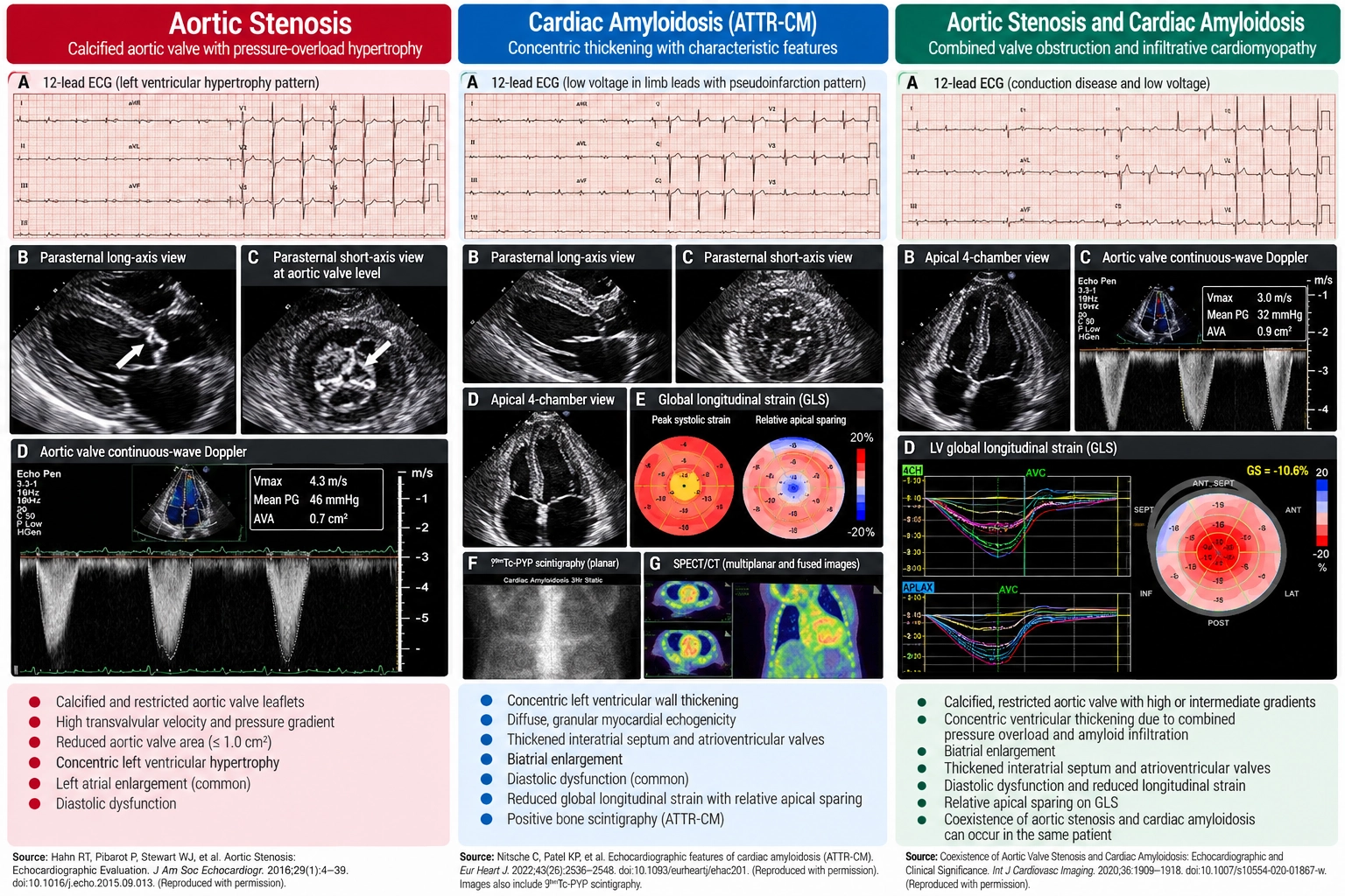
Figure 1. Echocardiographic and Multimodality Imaging Findings in Aortic Stenosis, Cardiac Amyloidosis and Coexisting AS–ATTR-CM. Aortic stenosis is characterized by restricted aortic valve opening, increased transvalvular gradients, and pressure-overload ventricular hypertrophy. Transthyretin amyloid cardiomyopathy (ATTR-CM) commonly produces increased ventricular wall thickness, diastolic dysfunction, and reduced global longitudinal strain (GLS), sometimes with relative apical sparing. When both conditions coexist, valvular obstruction and myocardial amyloid infiltration may contribute to ventricular dysfunction and low-flow, low-gradient physiology. These findings may overlap and require integrated clinical and multimodality assessment.
Low-Flow, Low-Gradient Aortic Stenosis: A Critical Diagnostic Challenge
One of the most important diagnostic difficulties occurs when severe aortic stenosis is associated with a low transvalvular pressure gradient.
In classical high-gradient severe AS, peak aortic velocity is at least 4.0 m/s or the mean gradient is at least 40 mmHg, generally with an aortic valve area of 1.0 cm² or less.
However, some patients have a calculated valve area of 1.0 cm² or less despite a mean gradient below 40 mmHg.
This discordance requires further assessment rather than automatically classifying the disease as severe.
What Is Low-Flow, Low-Gradient AS?
Low-flow, low-gradient aortic stenosis typically involves:
- aortic valve area of 1.0 cm² or less;
- mean transvalvular gradient below 40 mmHg; and
- stroke volume index of 35 mL/m² or less.
It may occur with reduced ejection fraction or with preserved ejection fraction, the latter often termed paradoxical low-flow, low-gradient AS.
Cardiac amyloidosis may contribute to reduced stroke volume through restrictive ventricular physiology, impaired longitudinal contraction, and reduced ventricular cavity size.
Consequently, a patient with both ATTR-CM and aortic stenosis may have a lower-than-expected pressure gradient despite clinically important valve obstruction.
How Is Discordant AS Severity Confirmed?
According to the 2025 ESC/EACTS valvular heart disease guidelines, evaluation may include:
- reassessment of Doppler measurements and left ventricular outflow tract dimensions;
- evaluation of stroke volume index and blood pressure;
- dobutamine stress echocardiography in selected patients, particularly with reduced LVEF;
- computed tomography assessment of aortic valve calcium; and
- integrated Heart Team evaluation when findings remain inconclusive.
Importantly, CT calcium thresholds require cautious interpretation in patients with concomitant amyloidosis or predominantly fibrotic valve disease (1).
Global Longitudinal Strain: Can Apical Sparing Identify Amyloidosis?
Global longitudinal strain measures longitudinal myocardial deformation using speckle-tracking echocardiography.
Both aortic stenosis and cardiac amyloidosis can reduce GLS even when ejection fraction remains preserved.
GLS in Cardiac Amyloidosis
Cardiac amyloidosis frequently produces more severe impairment in basal and mid-ventricular longitudinal strain, with relative preservation of apical strain.
This pattern is commonly called relative apical sparing.
GLS in Aortic Stenosis
Aortic stenosis can also cause regional and global strain abnormalities through pressure overload, hypertrophy, and fibrosis.
Some patients with severe aortic stenosis may demonstrate a relative apical-sparing pattern, even without confirmed amyloidosis.
Therefore, apical sparing should be considered a supportive clue rather than a definitive diagnostic sign.
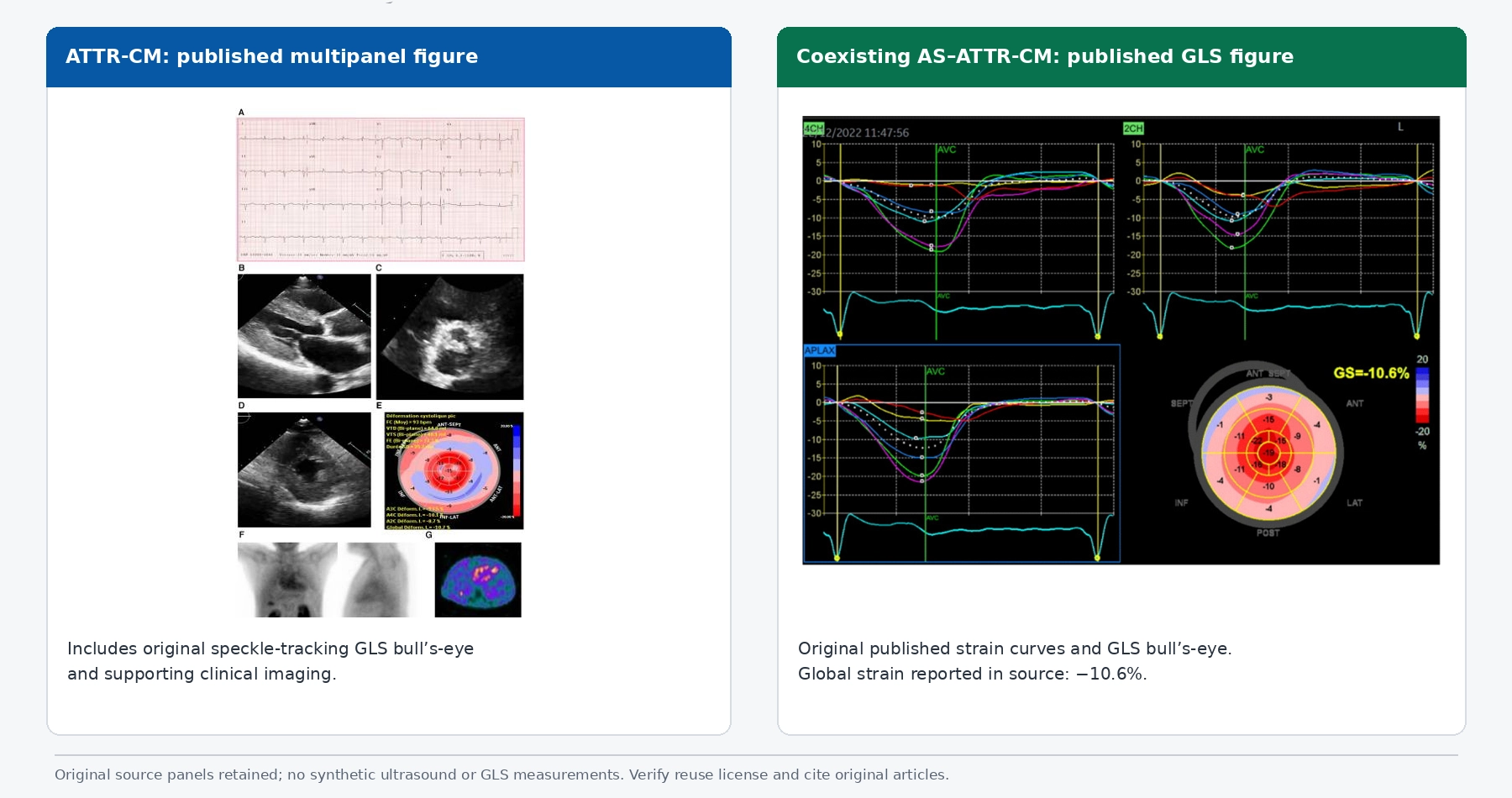
Figure 2. Global Longitudinal Strain in Cardiac Amyloidosis and Coexisting Aortic Stenosis. Published clinical images illustrating myocardial deformation patterns in transthyretin amyloid cardiomyopathy (ATTR-CM) and coexisting aortic stenosis with ATTR-CM. The left panel includes echocardiographic views and a GLS bull’s-eye map, while the right panel demonstrates longitudinal strain curves and a bull’s-eye map with a reported GLS of −10.6%. Relative apical sparing may support suspicion for cardiac amyloidosis; however, similar patterns can occur in aortic stenosis. GLS findings should therefore be interpreted alongside clinical assessment, valve hemodynamics and additional diagnostic investigations.
ECG Findings: The Voltage–Mass Discrepancy
ECG can provide useful information when clinicians investigate unexplained ventricular thickening.
Aortic stenosis may produce voltage criteria for left ventricular hypertrophy and associated repolarization abnormalities.
Cardiac amyloidosis may produce unexpectedly low QRS voltage relative to the degree of wall thickening seen on imaging.
Additional findings can include atrial fibrillation, conduction abnormalities, and pseudo-infarction patterns.
However, low voltage is not universal in ATTR-CM, and ECG voltage is influenced by several other clinical factors.
Therefore, the voltage–mass discrepancy is a useful warning sign, not a confirmatory test.
Cardiac MRI: Identifying Myocardial Infiltration
Cardiac magnetic resonance imaging provides tissue characterization that may help distinguish pressure-overload remodeling from infiltrative cardiomyopathy.
Late Gadolinium Enhancement
In aortic stenosis, CMR may demonstrate focal replacement fibrosis, often in a nonischemic mid-wall pattern. Diffuse interstitial fibrosis may also occur without conspicuous LGE.
In cardiac amyloidosis, LGE more frequently follows a diffuse subendocardial or transmural distribution.
Abnormal myocardial nulling may provide another clue to amyloid infiltration.
Native T1 Mapping and Extracellular Volume
Native T1 and extracellular volume fraction (ECV) can be elevated in both conditions.
In aortic stenosis, increased ECV commonly reflects myocardial fibrosis associated with chronic pressure overload.
In cardiac amyloidosis, amyloid deposition can produce substantial extracellular expansion and markedly elevated ECV.
However, measurements overlap. Native T1 values also vary according to scanner field strength, acquisition sequence, and institutional reference ranges.
Therefore, no single CMR value should be used as a universal diagnostic threshold for cardiac amyloidosis.
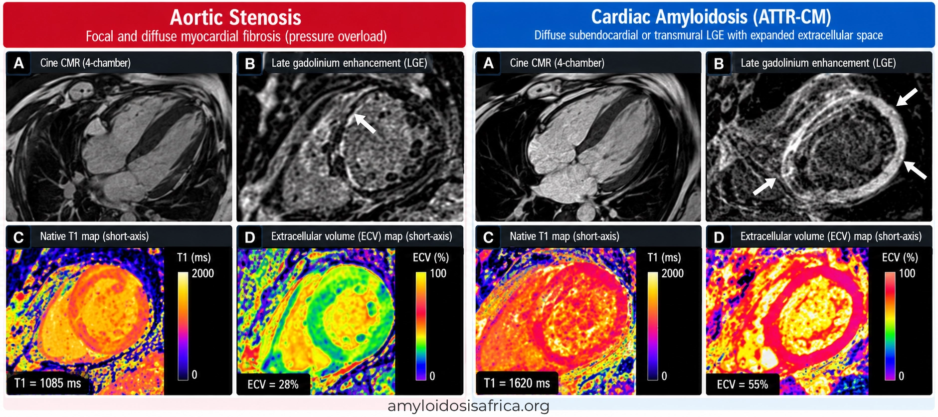
Figure 3. Cardiac MRI Differences Between Aortic Stenosis and Cardiac Amyloidosis. Cardiac magnetic resonance imaging illustrates differences in myocardial tissue characteristics between aortic stenosis and transthyretin amyloid cardiomyopathy (ATTR-CM). Aortic stenosis may produce focal mid-wall late gadolinium enhancement (LGE) and diffuse interstitial fibrosis, with variable increases in native T1 and extracellular volume (ECV). In contrast, cardiac amyloidosis frequently demonstrates diffuse subendocardial or transmural LGE, elevated native T1 and substantial ECV expansion, reflecting extracellular amyloid deposition. Although these findings provide valuable diagnostic clues, they may overlap and should be interpreted alongside clinical assessment and disease-specific investigations.
Clinical Red Flags for Coexisting ATTR-CM
Some findings should increase suspicion for ATTR-CM in a patient already diagnosed with aortic stenosis.
- older age with otherwise unexplained ventricular wall thickening;
- bilateral carpal tunnel syndrome;
- lumbar spinal stenosis;
- atraumatic biceps tendon rupture;
- peripheral or autonomic neuropathy;
- conduction disease or previous pacemaker implantation;
- unexpectedly low ECG voltage;
- heart failure disproportionate to the apparent severity of valve obstruction; and
- persistent heart failure symptoms after technically successful valve replacement.
These features should be interpreted collectively. None establishes ATTR-CM independently.
Comparison Table: Cardiac Amyloidosis vs Aortic Stenosis
| Feature | Cardiac Amyloidosis | Aortic Stenosis |
|---|---|---|
| Primary mechanism | Extracellular amyloid deposition | Obstruction of left ventricular outflow through the aortic valve |
| LV wall thickening | Common; infiltrative | Common; pressure-overload hypertrophy |
| Aortic valve obstruction | Not required for diagnosis | Defining feature |
| Diastolic dysfunction | Common | Common |
| GLS | Often reduced with relative apical sparing | May be reduced with overlapping regional patterns |
| ECG voltage | May be low relative to wall thickness | May show LVH voltage criteria |
| CMR LGE | Often diffuse subendocardial or transmural | May show focal mid-wall fibrosis |
| ECV | Frequently substantially elevated | May increase because of fibrosis |
| Diagnostic confirmation | Subtype-specific laboratory testing, scintigraphy or biopsy | Integrated echocardiographic and multimodality valve assessment |
| Treatment | Amyloid subtype-specific therapy | Valve intervention when indicated |
Diagnostic Approach When Both Diseases Are Suspected
Step 1: Establish Aortic Stenosis Severity
Assess peak aortic velocity, mean gradient, valve area, stroke volume index, ventricular function and valve morphology.
When measurements are discordant, investigate possible low-flow, low-gradient disease using the appropriate multimodality approach.
Step 2: Identify Amyloidosis Red Flags
Review ECG, echocardiographic features, GLS, cardiac biomarkers, extracardiac manifestations and relevant clinical history.
Step 3: Exclude AL Amyloidosis
When cardiac amyloidosis is suspected, perform a complete monoclonal protein screen:
- serum free light chains and kappa/lambda ratio;
- serum immunofixation; and
- urine immunofixation.
These investigations are essential because AL amyloidosis requires urgent specialist assessment.
Protein electrophoresis alone is insufficient to exclude AL amyloidosis.
Learn more about monoclonal protein testing.
Step 4: Evaluate for ATTR-CM
When monoclonal protein testing is negative, technetium-99m PYP, DPD, or HMDP scintigraphy with appropriate SPECT imaging may support a nonbiopsy diagnosis of ATTR-CM.
In the appropriate clinical context, grade 2 or 3 myocardial tracer uptake, together with a negative monoclonal protein screen, can establish ATTR-CM noninvasively (3).
However, grade 1 uptake is not diagnostic, and positive monoclonal testing requires additional evaluation because AL amyloidosis cannot be excluded by scintigraphy alone.
Biopsy with accurate amyloid typing may be required when findings are equivocal or discordant.
Read more about bone scintigraphy and amyloidosis biopsy.
Step 5: Perform TTR Genetic Testing
After ATTR amyloidosis is confirmed, TTR genetic testing distinguishes hereditary from wild-type disease and informs family counseling.
Does Cardiac Amyloidosis Change TAVR Decisions?
The coexistence of ATTR-CM and severe aortic stenosis creates an important management challenge.
Valve replacement relieves the mechanical obstruction caused by aortic stenosis. However, it does not remove myocardial amyloid deposits.
Therefore, patients with both conditions may continue to experience heart failure or other manifestations of amyloid cardiomyopathy after valve intervention.
Nevertheless, available evidence does not support automatically withholding TAVR solely because cardiac amyloidosis is present.
In the multicenter study by Nitsche and colleagues, patients with concomitant disease who underwent TAVR had better observed survival than those managed medically, although the observational design limits causal conclusions (2).
The 2025 ESC/EACTS guidelines also recognize that patients with concomitant severe AS and transthyretin cardiac amyloidosis usually benefit from appropriate valve intervention (1).
Decisions should be individualized by a multidisciplinary Heart Team, considering:
- confirmed aortic stenosis severity;
- symptom burden and functional status;
- cardiac amyloidosis subtype and stage;
- other comorbidities and frailty;
- anticipated procedural benefit;
- patient preferences; and
- availability of disease-specific treatment and follow-up.
What Happens if Heart Failure Persists After TAVR?
Persistent symptoms following successful valve replacement may have several explanations.
These include residual or unrelated valvular disease, atrial fibrillation, coronary artery disease, pulmonary hypertension, kidney dysfunction and pre-existing myocardial fibrosis.
However, undiagnosed cardiac amyloidosis should also be considered, particularly when ventricular thickening, conduction abnormalities or characteristic extracardiac red flags are present.
Recognizing ATTR-CM after TAVR may enable appropriate specialist referral and consideration of disease-modifying treatment.
Why the Correct Diagnosis Changes Treatment
Treatment of Aortic Stenosis
Management depends on valve severity, symptoms, ventricular function, anatomy, procedural risk and guideline recommendations.
Severe symptomatic AS generally requires valve intervention when appropriate, through transcatheter or surgical aortic valve replacement.
The optimal approach is determined through Heart Team assessment and shared decision-making.
Treatment of Cardiac Amyloidosis
Cardiac amyloidosis requires subtype-specific treatment.
For ATTR-CM, disease-modifying treatment may include approved transthyretin stabilizers or TTR-directed therapies, depending on the clinical indication and local regulatory availability.
For AL amyloidosis, treatment targets the underlying plasma-cell disorder and requires prompt specialist management.
Supportive heart failure treatment should be individualized because patients with amyloid cardiomyopathy may have limited tolerance of some cardiovascular medications.
Learn more about cardiac amyloidosis treatment and heart failure management.
Why This Distinction Matters in Africa
Across Africa, patients with valvular heart disease may face challenges in accessing advanced imaging, specialist evaluation and definitive interventions.
Meanwhile, the epidemiology of coexisting aortic stenosis and ATTR-CM remains insufficiently characterized in many African populations.
Most published prevalence estimates originate from older European and North American cohorts undergoing evaluation for TAVR.
Therefore, applying these figures directly to African populations would be inappropriate without local evidence.
Greater awareness among cardiologists, echocardiographers and heart failure specialists could improve recognition of cardiac amyloidosis in patients with apparently established valvular disease.
In addition, multicenter African research could help identify diagnostic gaps, characterize clinical presentations and develop practical referral pathways.
Learn more about cardiac amyloidosis in Africa.
Key Takeaways
- Aortic stenosis and cardiac amyloidosis can both produce ventricular wall thickening, diastolic dysfunction and heart failure.
- The conditions may coexist, especially in older adults with ATTR-CM.
- Low-flow, low-gradient AS requires careful confirmation of valve severity.
- Relative apical sparing on GLS is supportive but not specific for amyloidosis.
- CMR tissue characterization can help identify myocardial infiltration.
- A complete monoclonal protein screen is essential when amyloidosis is suspected.
- Bone scintigraphy can establish ATTR-CM noninvasively under defined diagnostic conditions.
- Coexisting cardiac amyloidosis does not automatically preclude TAVR.
- Both the valvular disease and amyloid cardiomyopathy may require treatment.
Frequently Asked Questions
Can aortic stenosis cause ventricular thickening similar to cardiac amyloidosis?
Yes. Aortic stenosis produces pressure-overload hypertrophy, while cardiac amyloidosis causes extracellular amyloid deposition. Both can increase ventricular wall thickness and impair diastolic function.
Can a patient have both aortic stenosis and cardiac amyloidosis?
Yes. Coexisting aortic stenosis and ATTR-CM are increasingly recognized in older adults, particularly among patients evaluated for TAVR.
How common is ATTR-CM in patients referred for TAVR?
Selected published cohorts have reported clinically relevant ATTR-CM in a proportion of older patients referred for TAVR. In one multicenter study, grade 2 or 3 bone scintigraphy uptake was identified in 7.9% of participants. Prevalence depends on the population and diagnostic approach.
What is low-flow, low-gradient aortic stenosis?
It describes aortic stenosis with discordant valve area and pressure-gradient measurements in the presence of reduced forward flow. Additional assessment is required to confirm whether the stenosis is truly severe.
Does apical sparing confirm cardiac amyloidosis?
No. Relative apical sparing can occur in other cardiac conditions, including aortic stenosis. It is a diagnostic clue rather than a definitive test.
Can cardiac MRI distinguish amyloidosis from aortic stenosis?
CMR can provide useful information about myocardial infiltration and fibrosis. However, imaging findings overlap, and CMR cannot independently establish the amyloid subtype.
Should patients with ATTR-CM undergo TAVR?
Patients with concomitant severe aortic stenosis and ATTR-CM may benefit from TAVR when valve intervention is otherwise indicated. Decisions require individualized Heart Team evaluation.
Does TAVR treat cardiac amyloidosis?
No. TAVR relieves aortic valve obstruction but does not remove myocardial amyloid deposits or treat the underlying protein disorder.
Should persistent heart failure after TAVR prompt amyloidosis testing?
Persistent heart failure has multiple possible causes. However, cardiac amyloidosis should be considered when clinical, ECG or imaging findings suggest myocardial infiltration.
References
- Praz F, Borger MA, Lanz J, et al. 2025 ESC/EACTS Guidelines for the management of valvular heart disease. Eur Heart J. 2025;46:4635–4736. doi:10.1093/eurheartj/ehaf194.
- Nitsche C, Scully PR, Patel KP, et al. Prevalence and Outcomes of Concomitant Aortic Stenosis and Cardiac Amyloidosis. J Am Coll Cardiol. 2021;77(2):128–139. doi:10.1016/j.jacc.2020.11.006.
- Gillmore JD, Maurer MS, Falk RH, et al. Nonbiopsy Diagnosis of Cardiac Transthyretin Amyloidosis. Circulation. 2016;133(24):2404–2412. doi:10.1161/CIRCULATIONAHA.116.021612.
- Garcia-Pavia P, Rapezzi C, Adler Y, et al. Diagnosis and treatment of cardiac amyloidosis: a position statement of the ESC Working Group on Myocardial and Pericardial Diseases. Eur Heart J. 2021;42(16):1554–1568. doi:10.1093/eurheartj/ehab072.
- Kittleson MM, Ruberg FL, Ambardekar AV, et al. 2023 ACC Expert Consensus Decision Pathway on Comprehensive Multidisciplinary Care for the Patient With Cardiac Amyloidosis. J Am Coll Cardiol. 2023;81(11):1076–1126. doi:10.1016/j.jacc.2022.11.022.
This article is intended for educational purposes and does not replace individualized medical advice or specialist evaluation. Aortic stenosis and cardiac amyloidosis may coexist, and their diagnostic findings can overlap. Decisions about imaging, amyloidosis testing, valve intervention and disease-specific treatment should follow current clinical guidelines and be individualized by qualified healthcare professionals.



Leave a Reply