



Pacemaker in Cardiac Amyloidosis: How It Works & When It’s Used
Pacemaker therapy in cardiac amyloidosis may become necessary when amyloid-related disease affects the heart’s electrical conduction system and causes clinically important bradycardia or heart block. A pacemaker does not treat the underlying amyloidosis. Instead, it helps maintain an adequate heart rate when the heart’s natural electrical system can no longer do so reliably.
Conduction disease is particularly common in transthyretin cardiac amyloidosis (ATTR-CM), although it can also occur in AL amyloidosis. Patients may develop sinus-node dysfunction, atrioventricular block, bundle-branch disease, or other conduction abnormalities as cardiac involvement progresses (1, 2).
However, a diagnosis of cardiac amyloidosis alone is not an indication for pacemaker implantation. The decision is based on the presence and severity of bradycardia or conduction disease, associated symptoms, expected pacing requirement, and the patient’s overall clinical condition.
Key principle: A pacemaker treats clinically important slow heart rhythms and conduction block. It does not remove amyloid from the heart or stop amyloid production.
Why Does Cardiac Amyloidosis Cause Conduction Disease?
Cardiac amyloidosis develops when amyloid fibrils accumulate within the myocardium and other cardiac structures. In ATTR-CM, the precursor protein is transthyretin. In AL cardiac amyloidosis, the precursor is an abnormal immunoglobulin light chain produced by a plasma cell or B-cell clone.
Amyloid-associated structural changes can involve the cardiac conduction system, including the sinus node, atrioventricular node, His bundle, bundle branches, and Purkinje network.
As a result, patients may develop:
- sinus bradycardia;
- sinus-node dysfunction;
- first-degree atrioventricular block;
- second-degree atrioventricular block;
- high-grade or complete heart block;
- right or left bundle-branch block; and
- other intraventricular conduction delays.
Electrical disease can progress over time. For this reason, expert guidance recommends close monitoring for clinically important conduction abnormalities and the possible need for permanent pacing (1).
Which Type of Amyloidosis Is Most Associated With Pacemaker Need?
Conduction-system disease is particularly well recognized in ATTR cardiac amyloidosis.
ATTR-CM may occur as hereditary ATTR amyloidosis or wild-type ATTR amyloidosis. Both forms can involve the cardiac conduction system.
Published observational data have shown that high-grade atrioventricular block requiring permanent pacing occurs in both hereditary and wild-type ATTR cardiac amyloidosis (3).
However, AL amyloidosis can also cause conduction abnormalities. Therefore, pacemaker decisions should depend on the patient’s electrical findings and symptoms rather than amyloid type alone.
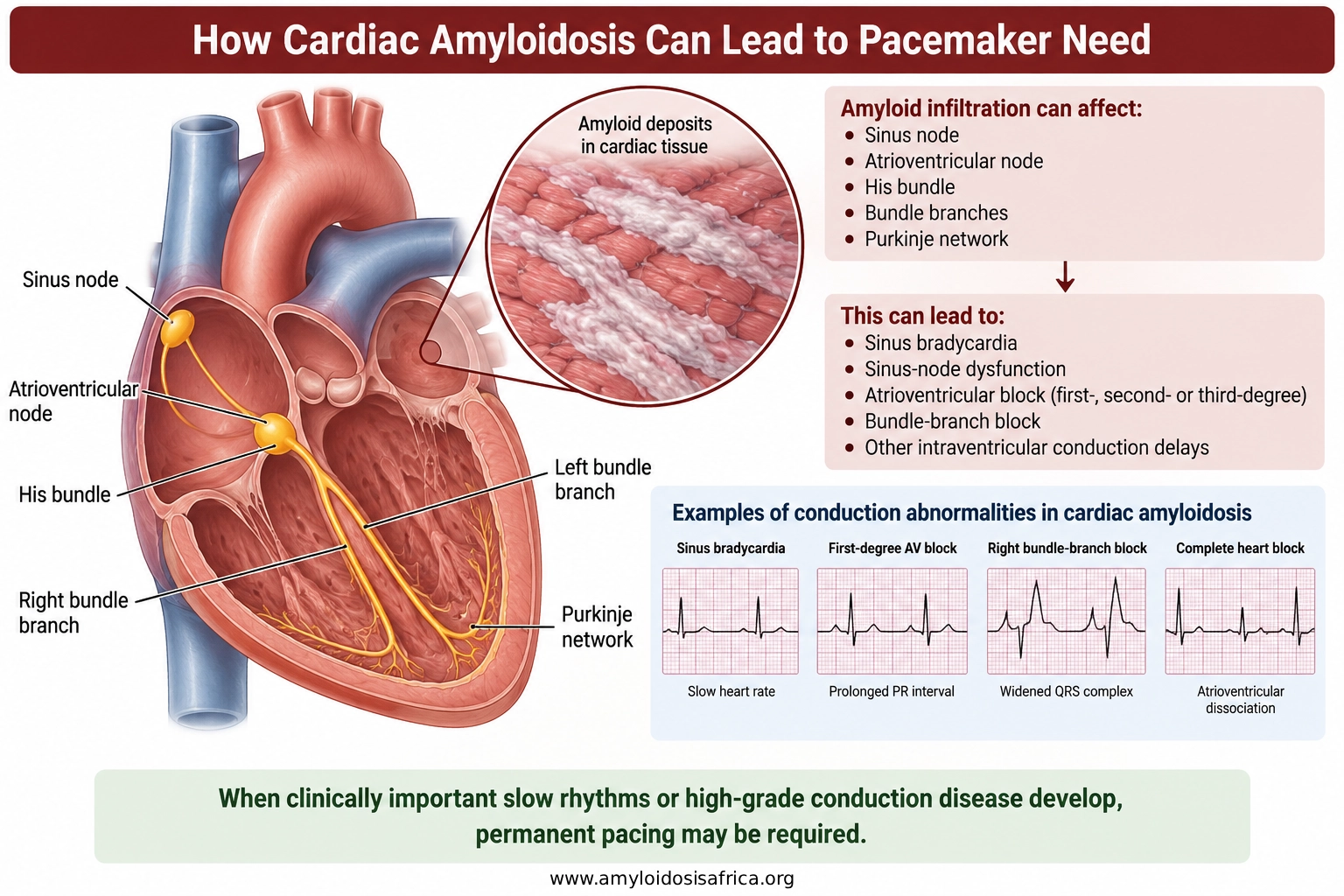
Figure 1. How Cardiac Amyloidosis Can Lead to Pacemaker Need. Amyloid-related involvement of the sinus node, atrioventricular node, His–Purkinje system, and bundle branches can lead to bradycardia or progressive conduction block. When clinically important slow rhythms or high-grade conduction disease develop, permanent pacing may be required.
What Is a Pacemaker?
A permanent pacemaker is a small electronic device that monitors the heart rhythm and delivers electrical impulses when the intrinsic heart rate becomes too slow.
A conventional pacemaker generally contains two main components:
- Pulse generator: a small device containing the battery and electronic circuitry, usually placed under the skin in the upper chest.
- Pacing lead or leads: insulated wires that pass through a vein into the heart and allow the pacemaker to sense the cardiac rhythm and deliver electrical impulses.
The exact device configuration depends on the patient’s rhythm disorder and clinical needs.
How Does a Pacemaker Work?
The pacemaker continuously monitors the heart’s electrical activity. If the heart rate falls below the programmed threshold or conduction between the atria and ventricles fails, the device can deliver a small electrical impulse to stimulate cardiac contraction.
Most patients do not feel routine pacing impulses.
Heart rate becomes too slow, or conduction fails
↓
Pacemaker detects the abnormal rhythm
↓
Electrical pacing impulse is delivered
↓
Heart contraction is triggered
↓
Pacemaker detects the abnormal rhythm
↓
Electrical pacing impulse is delivered
↓
Heart contraction is triggered
Single-Chamber Pacemaker
A single-chamber pacemaker usually has one lead placed in either the right atrium or right ventricle, depending on the pacing indication.
In patients with chronic atrial fibrillation and clinically significant bradycardia, for example, ventricular pacing may be sufficient in some situations.
Dual-Chamber Pacemaker
A dual-chamber pacemaker typically uses one lead in the right atrium and another in the right ventricle.
This configuration allows the device to coordinate atrial and ventricular activity more closely and may be appropriate for many patients with atrioventricular conduction disease.
Biventricular Pacing
Some patients require a more advanced pacing strategy known as cardiac resynchronization therapy (CRT). A CRT system generally provides pacing to both ventricles to improve ventricular electrical and mechanical synchrony.
CRT is not routinely required simply because a patient has amyloidosis. It may be considered when conventional CRT indications are present or when substantial ventricular pacing is expected, and the clinical team believes resynchronization may be advantageous.
When Is a Pacemaker Considered?
Pacemaker implantation is generally based on established pacing indications.
Clinical situations may include:
- symptomatic sinus-node dysfunction;
- clinically important bradycardia;
- high-grade atrioventricular block;
- complete heart block;
- selected second-degree AV block;
- clinically important pauses;
- tachycardia-bradycardia syndrome; and
- certain situations after AV-junction ablation.
The presence of cardiac amyloidosis can increase clinical concern because conduction disease may progress, but prophylactic pacemaker implantation is not routinely recommended in the absence of an established indication.
Important: An abnormal ECG does not automatically mean a pacemaker is required. The indication depends on the type and severity of conduction disease, symptoms, monitoring findings, and overall clinical context.
What Symptoms May Suggest Clinically Important Bradycardia?
Patients with cardiac amyloidosis may experience symptoms from bradycardia or conduction block, although similar symptoms can also result from heart failure, autonomic dysfunction, or other complications of amyloidosis.
Symptoms that may prompt rhythm assessment include:
- syncope;
- presyncope;
- dizziness;
- unexplained fatigue;
- exercise intolerance;
- weakness;
- shortness of breath; and
- episodes of unusually slow pulse.
Because symptoms are not specific, clinicians often combine symptom history with ECG and rhythm-monitoring data.
How Is Conduction Disease Evaluated?
Evaluation usually begins with a standard 12-lead ECG. Depending on the clinical situation, additional rhythm monitoring may be required.
Assessment may include:
- 12-lead ECG;
- 24- or 48-hour Holter monitoring;
- longer ambulatory rhythm monitoring;
- event monitoring;
- implantable loop recorder in selected patients;
- assessment of syncope or presyncope;
- echocardiography;
- cardiac MRI when clinically appropriate; and
- electrophysiology consultation when needed.
The clinician also evaluates heart-failure severity, atrial fibrillation, ventricular function, amyloid type, and extracardiac disease before selecting the most appropriate device strategy.
What ECG Changes May Occur Before Pacemaker Implantation?
Patients may show progressive conduction abnormalities before they develop high-grade heart block.
Examples include prolonged PR interval, bundle-branch block, widening of the QRS complex, and combinations of conduction defects.
However, no single ECG abnormality reliably determines exactly when a patient will require pacing. Serial clinical and electrocardiographic follow-up is therefore important.
How Is a Pacemaker Implanted?
Most conventional pacemakers are implanted through a minimally invasive transvenous procedure rather than open-heart surgery.
1. Preparation
The clinical team reviews the pacing indication, medications, anticoagulation, allergies, blood tests and relevant imaging.
During the procedure, ECG, blood pressure, and oxygen levels are monitored continuously. Local anesthesia is used at the implant site, usually together with sedation according to the patient’s clinical condition.
2. Creating the Pacemaker Pocket
A small incision is made in the upper chest, usually below the collarbone.
The operator creates a pocket beneath the skin or deeper tissues where the pulse generator will be placed.
3. Obtaining Venous Access
A vein is accessed in the upper chest region. Through this venous route, one or more pacing leads are advanced toward the heart.
4. Positioning the Leads
Under fluoroscopic guidance, pacing leads are positioned in the appropriate cardiac chamber or chambers.
For a dual-chamber pacemaker, one lead is commonly placed in the right atrium and another in the right ventricle.
The operator checks electrical sensing, pacing thresholds, and lead stability before completing implantation.
5. Connecting the Pulse Generator
The leads are connected to the pacemaker generator, which is then placed in the prepared pocket.
The system is tested and programmed according to the patient’s rhythm and pacing requirements.
6. Closing the Incision
Once the device is functioning appropriately, the incision is closed, and a dressing is applied.
The patient is then monitored during recovery.
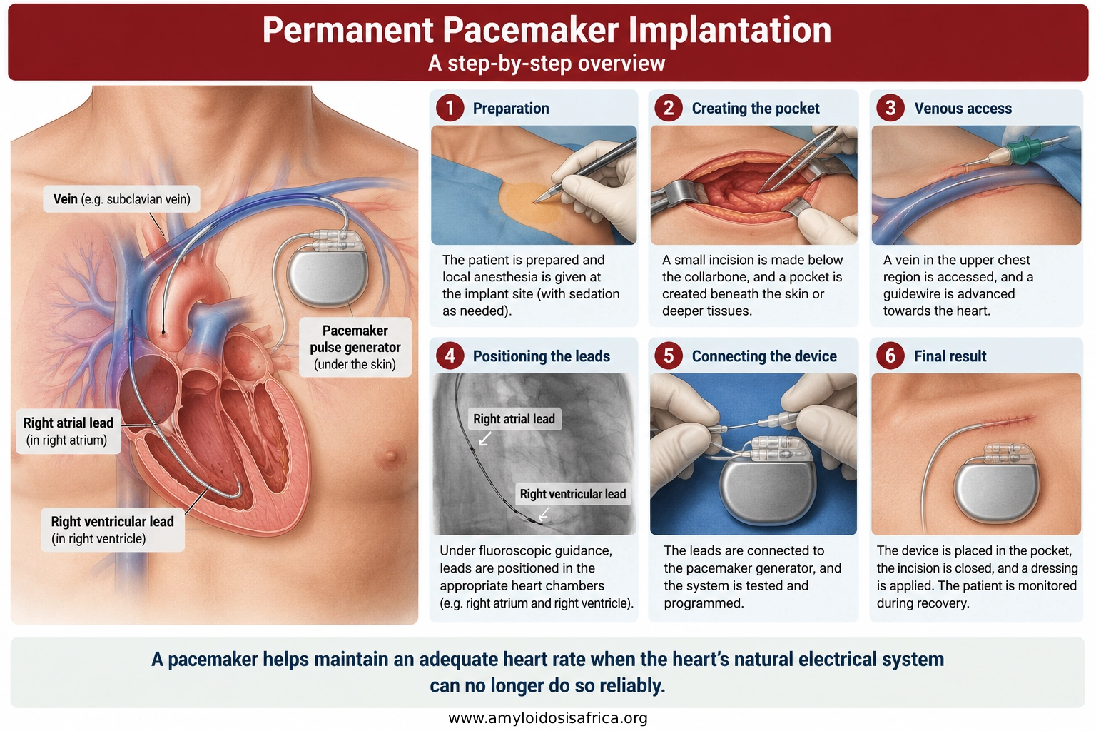
Figure 2. Permanent Pacemaker Implantation. During conventional transvenous pacemaker implantation, one or more leads are advanced through a vein into the heart and connected to a pulse generator placed beneath the skin in the upper chest. The device is tested and programmed before the incision is closed.
What Happens After Pacemaker Implantation?
After implantation, the clinical team checks the device and monitors for early complications.
Follow-up may include:
- device interrogation;
- assessment of sensing and pacing thresholds;
- wound inspection;
- ECG monitoring;
- chest imaging when clinically indicated;
- review of symptoms;
- assessment of percentage ventricular pacing; and
- instructions on wound care and activity.
Many patients can leave hospital within a short period after an uncomplicated procedure, although patients with advanced amyloidosis or significant comorbidities may require longer monitoring.
What Are the Risks of Pacemaker Implantation?
Pacemaker implantation is a common cardiac procedure, but complications can occur.
Potential risks include:
- bleeding or pocket hematoma;
- infection;
- pneumothorax;
- vascular injury;
- cardiac perforation or pericardial effusion;
- lead displacement;
- lead malfunction or fracture;
- device-pocket complications;
- need for lead revision; and
- future generator replacement when the battery approaches depletion.
The individual risk depends on the patient’s age, anatomy, medications, comorbidities and procedural complexity.
Why Does Right-Ventricular Pacing Burden Matter?
This is particularly important in cardiac amyloidosis.
Conventional right-ventricular pacing activates the ventricles in a different sequence from normal intrinsic conduction. If a large proportion of heartbeats are paced from the right ventricle, the resulting electrical dyssynchrony can adversely affect cardiac function in susceptible patients.
In a retrospective study of patients with ATTR cardiac amyloidosis and implanted devices, a right-ventricular pacing burden above 40% was associated with greater deterioration in left ventricular ejection fraction, mitral regurgitation and NYHA functional class compared with lower pacing burdens (4).
By contrast, patients receiving biventricular pacing showed more favorable changes in several clinical and echocardiographic measures.
These findings are observational and should not be interpreted as proof that every patient with cardiac amyloidosis and frequent pacing requires CRT. However, they highlight the importance of considering expected pacing burden when planning long-term device therapy.
Clinical consideration: When a patient with cardiac amyloidosis is expected to require substantial ventricular pacing, the electrophysiology and heart-failure teams may consider whether an alternative pacing strategy could reduce long-term ventricular dyssynchrony.
What Is the Difference Between Pacemaker and CRT?
| Feature | Conventional Pacemaker | CRT |
|---|---|---|
| Main purpose | Treat clinically important bradycardia or conduction block | Coordinate ventricular activation and reduce electrical dyssynchrony |
| Typical lead configuration | One or two leads, commonly involving the right atrium and/or right ventricle | Usually includes right-sided leads plus a lead positioned to pace the left ventricle through the coronary sinus |
| Role in amyloidosis | Used when standard pacing indications are present | May be considered when conventional CRT indications or substantial anticipated pacing are present |
| Amyloidosis-specific evidence | Pacemaker need is well recognized | Evidence remains limited and mainly observational |
| Treats amyloid deposition? | No | No |
What Is the Difference Between a Pacemaker and an ICD?
A pacemaker and an implantable cardioverter-defibrillator are not the same device, although some ICDs can also provide pacing.
| Feature | Pacemaker | ICD |
|---|---|---|
| Main function | Treat slow heart rhythms | Detect and treat selected dangerous ventricular tachyarrhythmias |
| Bradycardia pacing | Yes | Available in most transvenous ICD systems |
| Defibrillation shock | No | Yes |
| Typical indication | Clinically important bradycardia or conduction block | Selected patients at significant risk of ventricular tachycardia or ventricular fibrillation |
For more detail, see ICD implantation in cardiac amyloidosis.
Can Atrial Fibrillation Lead to Pacemaker Implantation?
Atrial fibrillation itself does not automatically require a pacemaker.
However, some patients develop slow ventricular rates, long pauses or tachycardia-bradycardia syndrome.
In selected patients with difficult rate control or refractory atrial fibrillation, AV-junction ablation followed by permanent pacing may also be considered as a later treatment strategy when appropriate.
What Is AV-Junction Ablation With Pacing?
AV-junction ablation intentionally interrupts electrical conduction between the atria and ventricles. After the procedure, the ventricles depend on permanent pacing.
This strategy may be considered in selected patients with atrial fibrillation when adequate rate control cannot be achieved using medication or other rhythm-management strategies.
Because the procedure creates pacemaker dependence, careful selection of the pacing strategy is particularly important.
How Does Amyloid Type Affect Pacemaker Management?
The indication for pacing is based primarily on the electrical abnormality rather than the amyloid subtype.
Nevertheless, amyloid type remains clinically important because ATTR and AL have different natural histories and disease-specific treatments.
Accurate diagnosis may require monoclonal protein testing, bone scintigraphy, genetic testing and, in selected situations, biopsy with amyloid typing.
Pacemaker implantation should therefore occur within a broader amyloidosis management plan rather than as an isolated intervention.
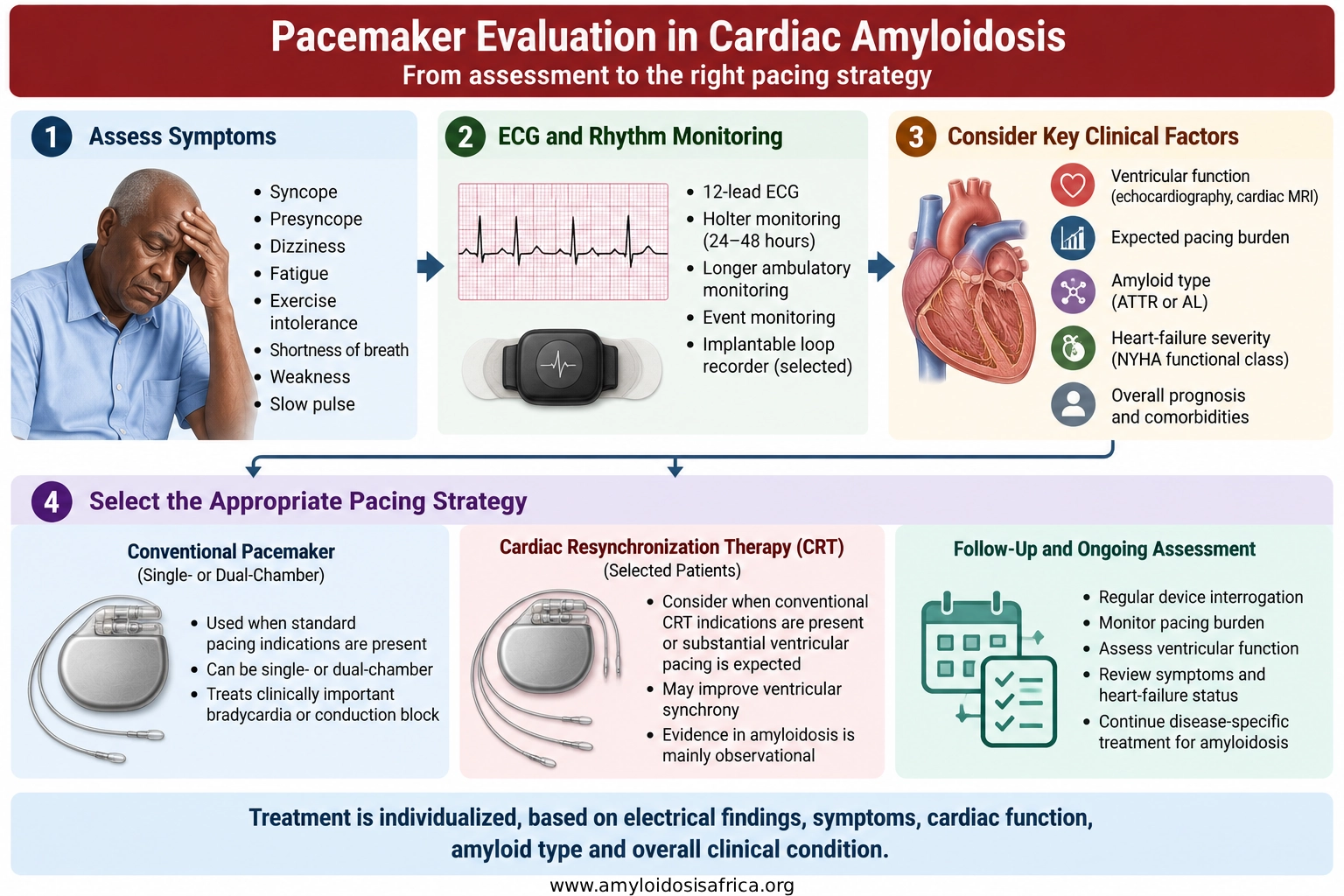
Figure 3. Pacemaker Evaluation in Cardiac Amyloidosis. Evaluation begins with symptoms, ECG and rhythm monitoring to identify clinically important bradycardia or conduction disease. Clinicians then consider ventricular function, expected pacing burden, amyloid type, heart-failure severity and overall prognosis before selecting a conventional pacemaker or, in selected patients, a more advanced pacing strategy.
Pacemaker Therapy Does Not Treat the Underlying Amyloidosis
A pacemaker can successfully correct clinically important slow heart rhythms, but it does not alter amyloid production or deposition.
Patients therefore continue to require appropriate cardiac amyloidosis treatment.
For ATTR-CM, treatment targets the transthyretin disease process. For AL amyloidosis, therapy targets the abnormal plasma-cell or B-cell clone producing amyloidogenic light chains.
Heart-failure management, atrial fibrillation treatment, anticoagulation when indicated and management of extracardiac disease remain necessary alongside pacemaker follow-up.
Follow-Up After Pacemaker Implantation
Permanent pacemakers require lifelong surveillance.
Device follow-up usually evaluates:
- battery status;
- lead integrity;
- pacing thresholds;
- electrical sensing;
- atrial and ventricular arrhythmias;
- percentage ventricular pacing;
- changes in heart-failure symptoms; and
- progression of cardiac amyloidosis.
Remote monitoring can allow device information to be transmitted between clinic visits where appropriate infrastructure is available.
What Happens When the Battery Runs Low?
Pacemaker batteries usually last for several years. The device estimates remaining battery life during routine interrogation.
When battery depletion approaches, the generator can usually be replaced through a smaller procedure while existing leads are retained if they remain functional.
This is called generator replacement rather than a completely new pacemaker implantation.
What If Conduction Disease Continues to Progress?
Progressive conduction disease may increase pacing requirements over time.
A patient who initially needs intermittent pacing can later become highly or completely pacemaker dependent.
This is why long-term monitoring of pacing burden and ventricular function is important in cardiac amyloidosis.
If substantial right-ventricular pacing is associated with worsening ventricular function or heart failure, clinicians may reassess whether a different pacing strategy is appropriate.
What About Advanced Heart Failure?
A pacemaker cannot reverse end-stage restrictive amyloid cardiomyopathy.
If heart failure progresses despite disease-specific therapy, careful volume management and appropriate device treatment, clinicians should reassess the overall treatment strategy.
Highly selected patients may be evaluated for heart transplantation in cardiac amyloidosis.
However, transplant assessment depends on amyloid type, extracardiac disease, frailty, functional status, control of the underlying disease process and expected long-term benefit.
Pacemaker Therapy and Cardiac Amyloidosis in Africa
Pacemaker implantation is available in several African cardiac centers, but access remains uneven across countries and regions.
Successful pacemaker programs require much more than the device itself. Sustainable care depends on:
- accurate diagnosis of conduction disease;
- appropriate patient selection;
- trained implanting cardiologists or electrophysiologists;
- sterile procedural facilities;
- fluoroscopy and imaging support;
- device programmers;
- management of procedural complications;
- long-term device interrogation;
- access to generator replacement; and
- reliable clinical follow-up.
Improving cardiac amyloidosis care in Africa therefore requires integrated diagnostic and referral pathways that link amyloidosis recognition, rhythm assessment, device services and disease-specific treatment.
More African data are also needed to define the prevalence of conduction disease, pacemaker implantation rates, pacing burden and long-term outcomes among patients with ATTR and AL cardiac amyloidosis.
Africa-focused priority: Expanding pacemaker access should occur alongside stronger cardiac amyloidosis diagnostic pathways, electrophysiology capacity, device follow-up systems and disease-specific treatment.
Key Takeaways
- Pacemakers treat clinically important slow heart rhythms and conduction disease in cardiac amyloidosis.
- Conduction abnormalities are particularly common in ATTR cardiac amyloidosis but can also occur in AL amyloidosis.
- Cardiac amyloidosis alone is not an indication for routine pacemaker implantation.
- Pacemaker implantation generally follows established pacing indications such as symptomatic bradycardia or high-grade AV block.
- Most conventional pacemakers are implanted through a transvenous procedure with a generator placed under the upper-chest skin.
- Patients may receive single-chamber, dual-chamber or more advanced pacing systems depending on their electrical and cardiac needs.
- A high right-ventricular pacing burden may adversely affect ventricular function in some patients with ATTR cardiac amyloidosis.
- CRT may be considered in selected patients with conventional indications or substantial anticipated pacing, but amyloidosis-specific evidence remains limited.
- Pacemaker therapy does not treat amyloid production or remove amyloid deposits.
- Lifelong device follow-up and continued amyloidosis-specific treatment remain essential.
Frequently Asked Questions
Do All Patients With Cardiac Amyloidosis Need a Pacemaker?
No. Pacemaker implantation is usually reserved for patients who develop a clinically important pacing indication such as symptomatic bradycardia or advanced conduction block.
Is Heart Block Common in Cardiac Amyloidosis?
Yes. Atrioventricular and His–Purkinje conduction disease are recognized manifestations of cardiac amyloidosis, particularly ATTR-CM.
Can a Pacemaker Prevent Sudden Cardiac Death?
A pacemaker prevents problems caused by excessively slow heart rhythms. It does not provide defibrillation for ventricular fibrillation or dangerous ventricular tachycardia unless the patient has a device with ICD functionality.
Is Pacemaker Implantation Open-Heart Surgery?
No. Most conventional pacemakers are implanted through a small upper-chest incision, with leads advanced through a vein into the heart.
Can a Pacemaker Treat Atrial Fibrillation?
A pacemaker does not eliminate atrial fibrillation. However, it can treat associated slow heart rates or pauses and may support selected strategies such as AV-junction ablation.
Can Pacemaker Pacing Weaken the Heart?
Frequent conventional right-ventricular pacing can cause ventricular dyssynchrony in some patients. Observational data in ATTR cardiac amyloidosis suggest that a high right-ventricular pacing burden may be associated with worsening ventricular function.
Does a Pacemaker Cure Cardiac Amyloidosis?
No. A pacemaker treats an electrical complication of the disease. Patients still require treatment directed at the underlying ATTR or AL amyloidosis.
How Long Does a Pacemaker Last?
Battery longevity varies according to device type and pacing requirement, but pacemaker generators commonly function for several years before replacement is required.
References
- Kittleson MM, Ruberg FL, Ambardekar AV, et al. 2023 ACC Expert Consensus Decision Pathway on Comprehensive Multidisciplinary Care for the Patient With Cardiac Amyloidosis. J Am Coll Cardiol. 2023;81(11):1076–1126. doi:10.1016/j.jacc.2022.11.022.
- Garcia-Pavia P, Rapezzi C, Adler Y, et al. Diagnosis and treatment of cardiac amyloidosis: a position statement of the ESC Working Group on Myocardial and Pericardial Diseases. Eur Heart J. 2021;42(16):1554–1568. doi:10.1093/eurheartj/ehab072.
- Donnellan E, Wazni OM, Saliba WI, et al. Prevalence, incidence, and impact on mortality of conduction system disease in transthyretin cardiac amyloidosis. Am J Cardiol. 2020;128:140–146. doi:10.1016/j.amjcard.2020.05.021.
- Donnellan E, Wazni OM, Saliba WI, et al. Cardiac devices in patients with transthyretin amyloidosis: impact on functional class, left ventricular function, mitral regurgitation, and mortality. J Cardiovasc Electrophysiol. 2019;30(11):2427–2432. doi:10.1111/jce.14180.
- Donnellan E, Wazni OM, Hanna M, Kanj M, Saliba WI, Jaber WA. Cardiac resynchronization therapy for transthyretin cardiac amyloidosis. J Am Heart Assoc. 2020;9(14):e017335. doi:10.1161/JAHA.120.017335.
- Rehorn MR, Loungani RS, Black-Maier E, et al. Cardiac implantable electronic devices: a window into the evolution of conduction disease in cardiac amyloidosis. JACC Clin Electrophysiol. 2020;6(9):1144–1154.
- Ruberg FL, Grogan M, Hanna M, Kelly JW, Maurer MS. Transthyretin amyloid cardiomyopathy: JACC State-of-the-Art Review. J Am Coll Cardiol. 2019;73(22):2872–2891. doi:10.1016/j.jacc.2019.04.003.
Medical Disclaimer
This article provides general educational information and does not replace individualized medical advice, diagnosis or treatment. Decisions regarding permanent pacemaker implantation or pacing strategy should be made by qualified cardiology and electrophysiology professionals after assessment of the individual patient’s conduction disease, symptoms, cardiac function, amyloid type, comorbidities and expected clinical benefit.

Leave a Reply